Equine Massage: A Practical Guide (31 page)
Read Equine Massage: A Practical Guide Online
Authors: Jean-Pierre Hourdebaigt

Kinesiology of the Horse
161
this leg movement. The lateral vastus muscle (point 3; figure 7.6) allows extension of the stifle, which in turn causes the hock and fetlock joints to extend. The biceps femoris, the gastrocnemius, and the deep flexor muscles (points 4, 5, and 6; figure 7.6) assist the flexion of the stifle and the fetlock.
All the muscles involved in the protraction of the hind limb are elongated during retraction and through their eccentric contraction ensure stability and smoothness of action.
Abduction
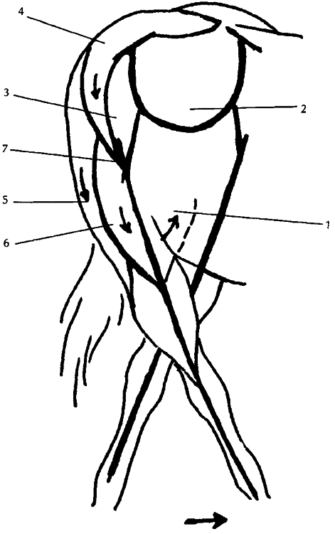
The muscles involved in the concentric contraction (which initiates abduction motion of the hind leg) are (see figure 7.7): 1. The medial gluteal muscle
2. The deep gluteal muscle
3. The superficial gluteal muscle
4. The biceps femoris muscle
5. The lateral vastus muscle
The elongated muscles in the abduction of the hind leg are: 7. The great adductor muscle group
These muscles attach along
the bones of the hind leg.Their
interplay will cause the abduction movement. The lateral
vastus muscle of the quadriceps
group (point 5; figure 7.7) pulls
the stifle laterally (outward). It
is assisted by the biceps femoris
and the tensor fasciae latae
muscles (points 4 and 6; figure
7.7), which pull on the femur
bone laterally (outward). The
antagonist muscles (points 7
and 8; figure 7.7), by their
eccentric contraction, contribute to the smoothness of
action.
7.7 Hind Leg Abduction
162
Equine Massage
Adduction
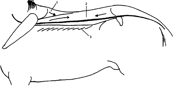
The muscles involved in the concentric contraction (which initiates adduction motion of the hind leg) are (see figure 7.8): 1. The great adductor muscle group
The elongated muscles in the adduction of the hind leg are: 3. The deep gluteal muscle
4. The superficial gluteal and the medial gluteal 5. The biceps femoris muscle
6. The lateral vastus muscle
These muscles attach along the bones of the hind leg. Their interplay will cause the adduction movement.The great adductor muscle group (point 1; figure 7.8) mostly causes this action by pulling the hind leg medially (inward). The antagonist muscles (points 3, 4, 5, 6, and 7; figure 7.8), by their eccentric contraction, contribute to the smoothness of action.
7.8 Hind Leg Adduction
Kinesiology of the Horse
163
The Vertebral Column
Besides protecting the spinal cord, the vertebral column provides a frame-like structure composed of strong bones, very thick ligaments, and muscles that bridge the anterior and posterior limbs.
Its role is to anchor strong muscle groups as well as to resist the downward effect of the center of gravity in mid-chest. In most equine disciplines, the vertebral column also supports the weight of a rider.
Extension
The agonist muscles responsible for the extension of the column are located above the vertebral column. This extensor muscle group is made of (see figure 7.9):
1. The spinalis dorsi muscle
2. The longissimus dorsi muscle
Flexion
The agonist muscles responsible for the flexion of the vertebral column are the abdominal muscle group and the intercostal muscles. The muscles associated with protraction, retraction, abduction, and adduction of the limbs have a secondary function, which is to assist with the flexion of the vertebral column.
Lateral Flexion
Lateral bending is not caused by any specific muscle. In fact, such bending is the result of a unilateral concentric contraction of
7.9 Back Extension
164
Equine Massage
either the flexor or extensor muscles of the spine. In this movement, the intervertebral muscles play a strong role. Running from one vertebra to the next, the intervertebral muscles are tiny muscles along the side of the vertebral column. The grand oblique muscle of the abdominal group, especially the internal oblique, is also an important player in this particular movement.
The Rib Cage
The pectoral muscles and the ventral serrate muscles play an important role in supporting and stabilizing the rib cage, or chest, in relation to the spine.The abdominal muscles assist lateral bending as well as support the rib cage. The intercostal muscles are responsible for the actual movement of the ribs. The diaphragm muscle is responsible for breathing.
The Neck
Muscles
Because the horse uses his head to counterbalance the weight in the rest of his body, the neck muscles play a very significant role in locomotion. Most obvious at the gallop, but also seen at the trot or walk, the downward swing of the head helps to lift the hind legs off the ground as the horse moves forward.
The neck muscles are:
2. The splenius (capitis and cervicis) muscle
3. The complexus muscle
5. The serrate muscle (cervical part)
6. The trapezius muscle (cervical part)
7. The rectus capitis ventralis muscle
8. The sternocephalicus muscle
9. The sternothyrohyoid and omohyoid muscle
10. The brachiocephalic muscle
11. The scalene muscle
These muscles are attached along the spine from the base of the skull, down the cervical vertebrae to the thoracic vertebrae, and to the upper ribs, and the scapulas. Their interplay will induce several different movements.
Kinesiology of the Horse
165
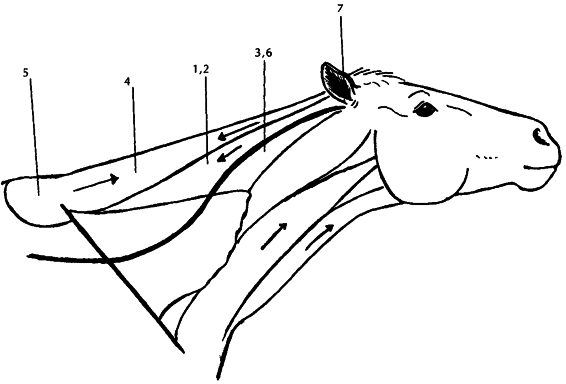
7.10 Neck Extension
Extension of the Neck
The muscles involved in the extension of the neck are (see figure 7.10):
1. The splenius muscle
3. The longissimus muscle of the neck
4. The trapezius muscle
5. The rhomboid muscle
6. The rectus capitis ventralis muscle
As these muscles contract, they cause the cervical section of the spine to arch in extension, bringing the head upwards.
All the muscles involved in the flexion of the neck are elongated during the extension movement, and through their eccentric contraction ensure stability and smoothness of action.
Flexion
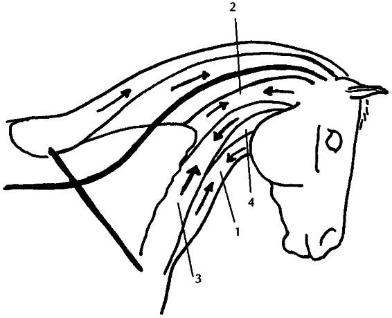
The neck muscles involved in the flexion of the neck are (see figure 7.11):
1. The sternocephalicus muscle
166
Equine Massage
7.11 Neck Flexion
3. The brachiocephalic muscle
As these muscles contract, they cause the cervical section of the spine to bend forward in flexion, bringing the head downward.
All the muscles involved in the extension of the neck are elongated during the flexion movement, and through their eccentric contraction ensure stability and smoothness of action.
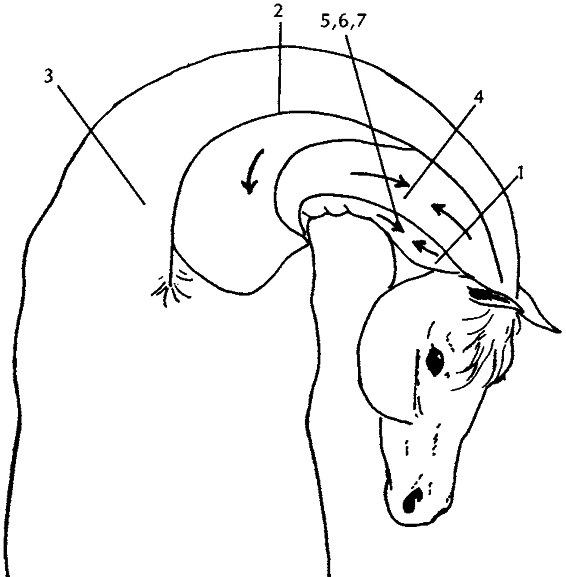
Lateral Flexion
The muscles involved in the lateral flexion of the neck are (see figure 7.12):
1. The rectus capitis muscle
2. The intervertebral muscles
3. The scalene muscle
4. The splenius cervicis muscle
5. The brachiocephalic muscle
6. The sternocephalicus muscle
The unilateral concentric contraction of these muscles to one side will cause the head and the cervical spinal column to move to the same side.
All the muscles involved on the opposite side of the neck are elongated, and through their eccentric contraction ensure stability and smoothness of action.
Kinesiology of the Horse
167
7.12 Lateral Neck Flexion
The Stay Mechanism of the
Horse
In the wild, horses cannot afford to spend much time lying down.
However, they do require adequate rest. Thus nature provides them with a mechanism that allows them to relax and rest while standing. This is known as the “stay” mechanism, which stabilizes both fore-and hind legs.The mechanism consists of ligaments and muscles that “lock” the main joints in the “stay” position.
Both front and hind lower legs have identical mechanisms based on the suspensory ligament and the superficial and deep digital flexor muscles whose tendons possess check ligaments.The rest of the structure is kept in an extended position by a system of muscles (see figure 7.13).
The ventral serrate muscle, both cervical and thoracic parts, mainly connects the foreleg to the body of the animal. The play between the cervical and thoracic parts keeps the scapula at a sloping angle, flexing the shoulder joint. The play between the biceps and the triceps muscles keeps the shoulder in extension.
The rest of the leg is well-aligned and the knee joint is prevented from bending forward by the lacertus fibrosus, an inelastic tendon that joins the biceps tendon and the radial carpal extensor muscle and tendon. Tension from the biceps is transmitted through this system to assist knee fixation.
