i bc27f85be50b71b1 (81 page)
Read i bc27f85be50b71b1 Online
Authors: Unknown

NE.RVOUS SYSTE.M
271
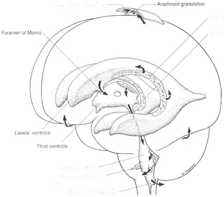
Supenor saglnal Sinus
Choroid plexus
Cerebral aqueduct
Fourth veolflcle
Foramen olluschka
Foramen 01 Magendle
Figure 4-3. The velltricular system of the brai". Arrows indicate the ciratlaliol1 of cerebrospinal fluid from the site of formatioll in the choroid plexus to the site of absorptioll ill the villi of the sagittal 5;11115. (\Vith permission from J
Bogouss/avsky, M Fisher /edsJ. Textbook of Neurology. Bostol1: Blitterworth
Heillemallll, 1998;656.)
\Vhen ventricular pressure is greater than venous pressure, CSF is
absorbed into the venous system via the arachnoid villi, capillary
walls of the pia mater, and lymphatics of the subarachnoid space near
the optic nerve.2
Blood-Brain Barrier
The blood-brain barrier is the physiologic mechanism responsible for
keeping toxins, such as amino acids, hormones, ions, and urea, from
altering neuronal firing of the brain. It readily allows water, oxygen,
carbon dioxide, glucose, some amino acids, and substances thar are
highly soluble in fat (e.g., alcohol, nicotine, and anesthetic agents) to
pass across the barrier,s,· The barrier consists of fused endothelial
cells on a basement membrane that is surrounded by astrocytic foot
extensions.· Substances must therefore pass through, rather than

272 ACUTE CARE HANDBOOK FOR PHYSICAL THERAPISTS
around, these cells. The blood-brain barrier is absent ncar the hypothalamus, pineal region, anterior third ventricle, and floor of the fourth ventricle.3
Central Brain Systems
The central brain systems are the reticular activating systcm and the
limbic system. The rcticular activating system is responsible for
human consciousness level and integrates the functions of the brain
stem with conical, cerebellar, thalamic, hypothalamic, and sensory
receptor functions.s
The limbic system is a complex interactive system, with primary
connections berween the cortex, hypothalamus, and sensory receptors.5 The limbic system is the emotional system, mediating cortical autonomic function of internal and external stimuli.
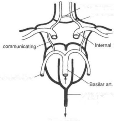
Circulation
The brain reccives blood from the internal carotid and vertebral arteries, which are linked together by the circle of Willis, as shown in Figure 4-4. Each vessel supplies blood to a certain part of the brain (Table 4-5). The circulation of the brain is discussed in tcrms of a sin-Ant communicating att.
AnI. cerebral art
Middle cerebral att
POSI.
carotid art
'".
POSI cerebral art
Greal cerebfal vein
(Galen)
CIrcle 01 WilNs
& associated veins
Figure 4-4. Schematic representation of the arterial circle of Willis and
accompanying veins. (Ant. anterior; art. artery; Post. posterior.) (\Vith
=
=
=
permission from £G Gonzalez, S) Meyers reds/. Downey and Darling's Physiological Basis of Rehabilitation Medicine (Jrd ed/. Boston: Butterworth
Heinemann, 2001 ;22.)


NERVOUS SYSTEM
273
Table 4-5. Blood Supply of the Major Areas of the Brain
Artery
Area of Perfusion
Anterior circulation
Internal carotid artery
The dura, optic tract, basal ganglia, midbrain,
(ICA)
uncus, lateral geniculate body, and tympanic
cavity. Ophthalmic branch supplies the eyes
and orbits.
External carotid artery
All structures external to the skull, the larynx,
(ECA)
and the thyroid.
Anterior cerebral artery
Medial and superior surface of frontal and
(ACA)
parietal lobes. Medial striate branch supplies anterior portion of the internal capsule,
optic chiasm and nerve, portions of the
hypothalamus, and basal ganglia.
Middle cerebral artery
Lateral surface of the frontal, parietal, and
(MCA)
occipital lobes, including the superior and
lareral surfaces of temporal lobes.
Posterior circulation
Vertebral artery
Medulla, dura of the posterior fossa, including
the falx cerebri and tentorium cerebelli.
Basilar artery
Pons and midbrain.
Posterior inferior
Posrerior and inferior surface of cerebellum.
cerebellar artery (PICA)
Anterior inferior
Anterior surface of the cerebellum, flocculus,
cerebellar artery (AICA)
and inferior vermis.
Superior cerebellar artery
Superior surface of cerebellum and vermis.
(SCA)
I)osterior cerebellar artery
Occipital lobe and medial and lateral surfaces
(PCA)
of the remporal lobes, thalamus, lareral geniculate bodies, hippocampus, and choroid
plexus of the third and lareral ventricles.
Sources: Data (rom CL Rumbaugh, A Wang, FY Tsai (cds). Cerebrovascular Disease
Imaging and Interventional Tre:nment Oprions. New York: Igaku-Shoin Medical Publishers, 1995; and KL Moore (cd). Clinically Oriented Anaromy (2nd cd). Baltimore: Williams & Wilkins, 1985.
gle vessel or by region (usually as the anterior or posterior circulation). There are several anastomotic systems of the cerebral vasculature that provide essential blood flow to the brain. Blood is drained from the brain through a series of venous sinuses. The superior sagittal sinus, with its associated lacunae and villi, is the primary drainage

274 ACUTE CARE HANDBOOK FOR PHYSICAL THERAI)ISTS
site. The superior sagittal sinus and sinuses located in the dura and
scalp then drain blood into the internal jugular vein for return to the
heart.
Spinal Cord
The spinal cord lies within the spinal column and extends from the
foramen magnum to the first lumbar vertebra, where ir forms the
conus medullaris and the cauda equina and attaches to the coccyx
via the filum terminale. Divided into the cervical, thoracic, and
lumbar portions, it is prorected by mechanisms similar ro those
supporting the brain. The spinal cord is composed of gray and
white matter and provides the pathway for the ascending and
descending tracts, as shown in cross section in Figure 4-5 and outlined in Table 4-6.
Peripheral Nervous System
The peripheral nervous sysrem consists of rhe cranial and spinal
nerves and the reflex system. The primary structures include
peripheral nerves, associated ganglia, and sensory receptors. There
are 12 pairs of CNs, each with a unique parhway and function
(sensory, motor, mixed, or autonomic). Thirty-one pairs of spinal
nerves (all mixed) exit the spinal cord to form distinct plexuses
(excepr T2 rhrough T12). The peripheral nerves of rhe upper and
lower extremities and thorax are listed in Tables 4-7 through 4-9,
and the dermatomal system is shown in Figure 4-6. The reflex system includes spinal, deep tendon, stretch, and superficial reflexes and protective responses.
Autonomic Nervous System
The portion of the peripheral nervous system thar innervates glands
and cardiac and smooth muscle is the autonomic nervous system.
The parasympathetic division is activated in time of rest, whereas
the sympathetic division is activated in times of work or "fight or
flight" situations. The two divisions work closely togerher, wirh dual
innervation of most organs, to ensure homeostasis.