Stretching Anatomy-2nd Edition (20 page)
Read Stretching Anatomy-2nd Edition Online
Authors: Arnold Nelson,Jouko Kokkonen
Tags: #Science, #Life Sciences, #Human Anatomy & Physiology

Stretch Notes
Injuries to the quadriceps muscles usually occur during an activity such as sprinting, jumping, or kicking, especially when the muscles are tight or unprepared for activity. This is yet another effective method of stretching the front thigh muscles. Although slightly more difficult than the beginner seated knee extensor stretch, this stretch still falls within the advanced beginner or intermediate category.
Because you perform this stretch while in a relaxed position, you have maximum control over the amount of stretch to the quadriceps muscles. In other words, this stretch allows you to concentrate solely on these thigh muscles while letting other muscles be as relaxed as possible.
Slowly pull the ankle in a more backward rather than upward direction while making sure the hips are also moving forward. Concentration should be greater on the forward hip movement than on the knee flexion (pulling the ankle toward the buttocks). As in any quadriceps muscle stretch, take extra care to prevent strain on the knee structure by overflexing the knee.
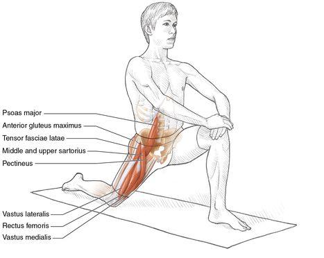
Advanced Kneeling Knee Extensor Stretch
Safety tip Do not attempt this stretch until you have moved past the beginner and intermediate knee extensor stretches.
Execution
- Step forward with the left leg, and bend the knee at about a 90-degree angle.
- Keep the left knee positioned above the left ankle.
- Extend the right leg behind the torso, and touch the floor with the right knee. The lower right leg lies on the floor.
- Hold on to an object or place the hands on the left knee to maintain balance.
- Move the hips forward, pushing the left knee in front of the left ankle and dorsiflexing that ankle.
- Repeat this stretch for the opposite leg.
Muscles Stretched
- Most-stretched muscles:
Right vastus medialis, right vastus intermedius, right vastus lateralis, middle and upper right sartorius, right rectus femoris, right psoas major, right iliacus, right tensor fasciae latae - Less-stretched muscles:
Right pectineus, anterior right gluteus maximus
Stretch Notes
The advanced kneeling knee extensor stretch is the quadriceps stretch most commonly used by athletes and nonathletes alike. Most people tend to have stronger but less flexible quadriceps muscles than hamstring muscles because of the tendency to stretch the hamstrings much more than the quadriceps. This creates an imbalance of strength and flexibility between the two muscle groups. To correct this imbalance, more emphasis needs to be placed on routinely stretching the quadriceps muscles.
When the right knee is extended behind the torso onto the floor, try to have a soft surface underneath the knee. This could be an exercise mat, grass, or even a pillow. This will minimize discomfort to the knee. When you move slowly into the stretched position, keep the left knee pointing forward. Do not let the left knee point to either side or let the right knee move along the surface of the floor. While the hips are placed in the forward direction, arching the back can increase the stretch on the muscles. This would stretch not only the quadriceps muscles but also the hip flexor muscles located in front of the pelvic area.
Advanced Supported Standing Knee Extensor Stretch

Execution
- Stand with the back toward a padded table, bed, or soft platform that is below the height of the hips.
- Balance your weight on the right leg, and bend the knee slightly.
- Bend the left knee, and prop the left ankle on the rear support surface.
- Place both hands on the rear support surface 6 to 12 inches (15 to 30 cm) behind the buttocks.
- Move the torso back slowly so that the heel of the left foot touches the buttocks. Make sure the ankle and knee are comfortable.
- Push the hips forward and simultaneously arch the back by bending the shoulders toward the buttocks.
- Repeat this stretch for the opposite leg.
Muscles Stretched
- Most-stretched muscles:
Left vastus medialis, left vastus intermedius, left vastus lateralis, middle and upper right sartorius, left rectus femoris, left psoas major, left iliacus, left tensor fasciae latae - Less-stretched muscles:
Left pectineus, anterior left gluteus medius
Stretch Notes
Knee stiffness can lead to injuries of the knee and of the quadriceps muscles and tendons. This is the most advanced stretch for the quadriceps muscles, and you must take extra care when attempting it. Because of the increased possibility of hyperflexing the knee, use this stretch only if you have very flexible muscles. By adhering to the following safety precautions, you can execute this stretch safely without injury.
While pulling the ankle slowly in a more backward than upward direction, concentrate on making sure your hips also move forward. This dual action stretches the hip flexor muscles located in front of the pelvic region as well as the quadriceps muscles. If you are experiencing soreness or tightness of either the lateral (outer) or medial (inner) side of the front thigh, consider placing most of the stretch emphasis on the medial muscles (vastus medialis and pectineus) by rotating the upper body away from the medial muscles (rotate the right side clockwise) when bending backward. To place most of the stretch emphasis on the lateral muscles (vastus lateralis and tensor fasciae latae), rotate the upper body away from the lateral muscles (rotate the right side counterclockwise) when bending backward.
For optimal results, it is important to brace both hands on the surface supporting the back. In addition, you should move your hips forward while carefully arching your back. This enables you to better control the amount of stretch being put on these muscles. Following these procedures maximizes stretch to the quadriceps muscles as well as to the hip flexor muscles located in front of the pelvic area. Yet another precaution for safety as well for comfort is having the ankle be up against the padded support behind you.
You might also consider moving the dorsal (top) part of the foot down to the padded support. This would bring additional benefits from the total stretch, because you also stretch the muscles in the anterior (front) part of the tibia bone in the lower leg. This is a powerful combination of multiple stretches.
In this stretch you are also able to change your trunk position, thus stretching the medial or lateral side of the thigh if you move your trunk in either a lateral (outer) or medial (inner) direction.
Chapter 7
Feet and Calves
The skeletal structure of the lower leg and foot is made up of the long tibia and fibula bones found in the lower leg and the small foot bones called tarsals, metatarsals, and phalanges. These bones form numerous joints. The most important is the ankle joint, located between the tibia bone of the leg and the talus of the foot. This joint is a hinge joint, and it is involved with the major joint movements of plantar flexion (toes point down) and dorsiflexion (toes point up).
The other major joints found between each of the tarsal and metatarsal bones are gliding joints. They allow more limited movements of the foot. When several of these gliding joints are working together in the foot, a much broader range of movement is achieved compared with the movement produced by a single gliding joint working alone. Thus, multiple-joint movements allow for foot eversion (sole turned out) and inversion (sole turned in).
The joints that allow the most freedom of movement of the foot are the condyloid joints, located between the metatarsal bones and the phalanges. Condyloid joints allow the movements of flexion, extension, abduction, adduction, and circumduction of the toes. Finally, the joints that allow for flexion and extension of the toes are the hinge joints between the phalanges.
Without the ligaments and connective tissues found in the lower leg and foot, joint movement and muscle function would be greatly compromised. The joints in the foot are connected to each other by many ligaments. The largest ligament in this area is the deltoid ligament, or ankle medial collateral ligament. It is composed of four segments that connect the tibia to the talus, calcaneus, and navicular bones. Opposite the deltoid ligament is the ankle lateral collateral ligament, which is composed of three segments that connect the fibula to the talus and calcaneus bones. Since the deltoid ligament is much stronger than the ankle lateral collateral ligament, and the tibia is longer than the fibula, the ankle is predisposed toward inversion (turning in).
Retinacula are another type of connective tissue located in the lower leg that secure many of the muscle–tendon units. This support allows these muscles to work harder, stronger, and more efficiently. The superior and inferior retinacula in the dorsal (top) area of the foot hold down all the tendons of the extensor muscles. On the lower lateral side of the foot, the peroneal retinaculum holds down the tendons of the peroneus longus and peroneus brevis muscles. The flexor retinaculum on the medial side of the ankle holds down the tendons of the flexor digitorum longus, flexor hallucis longus, and posterior tibialis muscles.
The final noteworthy connective tissue is the plantar fascia. The plantar fascia is a broad, thick connective tissue that supports the arch on the bottom of the foot. It spans the area between the tuberosity of the calcaneus and the heads of the metatarsal bones.
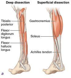
The muscles that move the ankle and toes are located primarily in the lower leg (
figure 7.1
); these muscles have tendons that are as long as or longer than the muscles. The dominant tendon is the Achilles tendon, which is shared by the gastrocnemius, plantaris, and soleus. The gastrocnemius and soleus muscles are the prime plantar flexors and are assisted by the plantaris and tibialis posterior as well as two toe flexor muscles, flexor digitorum longus and flexor hallucis longus. Located on the outer (lateral) side of the calf is another group of three muscles—peroneus longus, peroneus brevis, and peroneus tertius—which are used in everting the foot. Additionally, the peroneus longus and peroneus brevis plantar flex the ankle.
Three anterior calf muscles (tibialis anterior, extensor hallucis longus, and extensor digitorum longus) dorsiflex the ankle as well as move the foot and toes. The extensor digitorum brevis, dorsal interosseous, and extensor hallucis brevis muscles are located on the dorsal (top) side of the foot and extend the toes. The muscles on the plantar (sole) side of the foot, the flexor digitorum brevis, quadratus plantae, flexor hallucis brevis, flexor digiti minimi, abductor hallucis, abductor digiti minimi, plantar interosseous, and lumbricales, are used to flex and spread the toes.
Figure 7.1
Calf and foot muscles:
(a)
posterior;
(b)
anterior.