The Articulate Mammal (13 page)
Read The Articulate Mammal Online
Authors: Jean Aitchison

Although most neurologists agree that language is mainly restricted to one hemisphere, further localization of speech is still controversial. A basic difficulty is that until recently all the evidence available was derived from braindamaged patients. And injured brains may not be representative of normal ones. After a stroke or other injury the damage is rarely localized. A wound usually creates a blockage, causing a shortage of blood in the area beyond it, and a build-up of pressure behind it. So detailed correlations of wounds with speech defects cannot often be made, especially as a wound in one place may trigger off severe speech problems in one person, but only marginally affect the speech of another. This suggests to some neurologists that speech can be ‘re-located’ away from the damaged area – it has (controversially) been suggested that there are ‘reserve’ speech areas which are kept for use in emergencies. This creates an extremely complex picture. Like a ghost, speech drifts away to another area just as you think you have located it. But these problems have not deterred neurologists – and some progress has been made.
Until fairly recently, observation and experiment were the two main methods of investigation. Observation depended on unfortunate accidents and post-mortems. A man called Phineas Gage had an accident in 1847 in which a four-foot (over a metre long) iron bar struck and entered the front left-hand section of his head, then exited through the top. Gage kept the bar as a souvenir, until his death, twenty years later. The bar and skull are now preserved in a museum at the Harvard Medical School. Although Gage’s personality changed for the worse – he became unreliable and unpredictable – his language was unaffected. This suggests that the front part of the brain is not crucially involved in language. Conversely, a French surgeon named Broca noted at a post-mortem in 1861 that two patients who had had severe speech defects (one could only say
tan
and
sacré nom de Dieu
) had significant damage to an area just in front of, and slightly above, the left ear – which suggested that this area, now named ‘Broca’s area’, is important for speech.
The experimental method was pioneered in the 1950s by two Canadian surgeons, Penfield and Roberts (1959). They were primarily concerned with removing abnormally functioning cells from the brains of epileptics. But before doing this they had to check that they were not destroying cells involved in speech. So, with the patients fully conscious, they carefully opened the skull, and applied a minute electric current to different parts of the exposed brain. Electrical stimulation of this type normally causes temporary interference. So if the area which controls leg movement is stimulated, the patient is unable to move his or her leg. If the area controlling speech production is involved, the patient is briefly unable to speak.

There are obvious disadvantages in this method. Only the surface of the brain was examined, and no attempt was made to probe what was happening at a deeper level. The brain is not normally exposed to air or electric shocks, so the results may be quite unrepresentative. But in spite of the problems involved, certain outline facts became clear long ago.
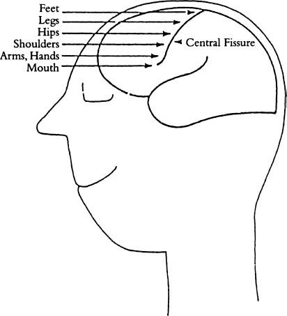
First of all, it was possible to distinguish the area of the brain which is involved in the actual articulation of speech. The so-called ‘primary somatic motor area’ controls all voluntary bodily movements and is situated just in front of a deep crack or ‘fissure’ running down from the top of the brain. The control for different parts of the body works upside down: control of the feet
and legs is near the top of the head, and control of the face and mouth is further down.
The bodily control system in animals works in much the same way – but there is one major difference. In humans, a disproportionate amount of space is allotted to the area controlling the hands and mouth.
But the sections of the brain involved in the actual articulation of speech seem to be partly distinct from those involved in its planning and comprehension. Where are these planning and comprehension areas? Experts disagree. Nevertheless, perhaps the majority of neurologists agree that some areas of the brain are statistically likely to be involved in speech planning and comprehension. Two areas seem to be particularly relevant: the neighbourhood of
Broca’s area
(in front of and just above the left ear); and the region around and under the left ear, which is sometimes called
Wernicke’s area
after the neurologist who first suggested this area was important for speech (in 1874). Damage to Wernicke’s area often destroys speech comprehension, and damage to Broca’s frequently hinders speech production – though this is something of an over-simplification, since serious damage to either area usually harms all aspects of speech (Mackay
et al.
1987).

Particularly puzzling are cases of damage to Broca’s or Wernicke’s area where the patient suffers no language disorder. Conversely, someone’s speech may be badly affected by a brain injury, even though this does not apparently involve the ‘language areas’. There may simply be more variation in the location of brain areas than in the position of the heart or liver. A particular function may be:
narrowly localized in an individual in a particular area … localized equally narrowly in another area in another individual, and carried out in a much larger area … in the third. The only constraint seems to be that core language processes are accomplished in this area of neocortex.
(Caplan 1988: 248)
A further problem is that neurologists do not necessarily agree on the exact location of Broca’s and Wernicke’s areas, though the boundaries are more contentious than the central regions (Stowe
et al
. 2005). In addition there are deeper brain interconnections about which little is known.
Comparisons with the brains of other primates, incidentally, show that humans have a disproportionately large area at the front of the brain, sometimes referred to as the ‘prefrontal cortex’, though it is unclear how much of this involves language, and how much more general interconnections.
Luckily, brain scans can now supplement our information. From the 1970s onward, these have moved forward in leaps and bounds. First, and prior to ‘proper’ scans was the EEG (electroencephalograph) which showed the numerous electrical impulses in the brain, and the general state of alertness of a patient, but was unable to provide precise mappings. Then came so-called CT or CAT scans, short for ‘X-ray computed tomography’. The tissues within the brain (and the body) differ in density, so a tumour (for example) might appear as an extra dense portion, and these differing densities showed up on the X-rays.
Next PET scans were developed, short for ‘positron emission tomography’. These recorded blood flow. Blood surges in the brain when someone uses language, just as extra blood is pumped into the arms and hands when someone plays the piano. Radioactive water was injected into a vein in the arm. In just over a minute, the water accumulated in the brain, and could show an image of the blood flow in progressively more difficult tasks. In one experiment, subjects were first asked to look at something simple, such as a small cross on a screen, and the blood flow was measured. Then, some English nouns were shown or spoken. As a next stage, the subjects were asked to speak the word they saw or heard. Finally, they were asked to say out loud a verb suitable to the noun: for example, if they had heard the word HAMMER, then HIT might be appropriate (Posner and Raichle 1994).
The results showed strong differences between the various tasks. Simply repeating involved only the areas of the brain which dealt with physical movement. But both Broca’s and Wernicke’s areas became active when subjects consciously accessed word meaning and chose a response. In short, comprehension and production cannot be split apart in the way it was once assumed. In production, selecting a verb was the most complex task, and involved several areas – though with practice, the activity grew less, and became more like that of nouns. So practice not only makes it all easier, but actually changes the way the brain organizes itself. In another experiment, subjects were presented with lists of verbs, and asked to provide the past tense. Regular past tenses such as CLIMBED, WISHED showed different blood flow patterns from irregular ones such as CAUGHT, HID (Jaeger
et al.
1996).
The brain, it appears, relies on tactics similar to those used by a sprinter’s muscles, with an increase in oxygen in any area where neurons show extra activity – though a basic problem is the tremendous amount happening at any one moment. Pinpointing only the activity relevant to speech is difficult, especially as deeper connections are turning out to be as important as those near the surface. However, techniques are improving all the time. These enable the ebb and flow in blood vessels to be monitored continuously.
More recently, attention has been directed particularly towards ERPs ‘event related potentials’, and MRI ‘magnetic resonance imaging’. These techniques are non-invasive, in that nothing needs to be injected into the body, which can simply be scanned. They are therefore potentially safer, and can be used with a wider range of people.
ERPs monitor electrical activity in the brain, following some stimulus (an ‘event’) such as reading a sentence. Electrodes are placed on the scalp, and the reaction, the ERP (‘event related potential’) is measured. The brain responds differently to syntactic and semantic ill-formedness, for example, showing that a division between the two has some type of ‘reality’ for speakers of English (Kutas and van Petten 1994).
MRI (‘magnetic resonance imaging’) exploits the finding that human heads and bodies contain hydrogen atoms which can be (temporarily and safely) re-aligned by means of the MRI machine’s magnetic field. Images of the brain are produced by taking photos of cross-sectional ‘slices’. These are far clearer and more precise than any previous attempts at picturing the brain. They confirm that a huge amount of activity takes place continuously.
The brain is therefore like an ever-bubbling cauldron, seething non-stop. Neurons are organized into complex networks: ‘The language areas may be understood as zones in which neurons participating in language-related cell assemblies cluster to a much higher degree than in other areas’ (Müller 1996: 629). But connections matter quite as much as locations, with far more buzzing between areas than was previously realized. So
connectionism
is the
general name for this type of theory about how the mind works, largely inspired by all this work on the brain. Multiple parallel links are turning out to be the norm in any mental activity, and especially in language. ‘Mental operations appear to be localized, but performance of a complex task requires an integrated ensemble of brain regions’ (Fiez
et al.
1992: 169).
PATTING ONE’S HEAD AND RUBBING THE STOMACH
As all this new work confirms, a type of biological adaptation which is not so immediately obvious – but which is on second sight quite amazing – is the ‘multiplicity of integrative processes’ (Lashley 1951) which take place in speech production and comprehension.
In some areas of activity it is extremely difficult to do more than one thing at once. As schoolchildren discover, it is extraordinarily hard to pat one’s head and rub one’s stomach at the same time. If you also try to swing your tongue from side to side, and cross and uncross your legs, as well as patting your head and rubbing your stomach, the whole exercise becomes impossible. The occasional juggler might be able to balance a beer bottle on his nose, twizzle a hoop on his ankle and keep seven plates aloft with his hands – but he is likely to have spent a lifetime practising such antics. And the exceptional nature of these activities is shown by the fact that he can earn vast sums of money displaying his skills.
Yet speech depends on the simultaneous integration of a remarkable number of processes, and in many respects what is going on is considerably more complex than the juggler’s manoeuvres with his beer bottle, plates and hoop.
In speech, three processes, at the very least, are taking place simultaneously: first, sounds are actually being uttered; second, phrases are being activated in their phonetic form ready for use; third, the rest of the sentence is being planned. And each of these processes is possibly more complicated than appears at first sight. The complexities involved in actually pronouncing words are not immediately apparent. One might assume that in uttering a word such as GEESE one first utters a G-sound, then an EE-sound, then an S–sound in that order. But the process is much more involved.
