Anatomies: A Cultural History of the Human Body (16 page)
Read Anatomies: A Cultural History of the Human Body Online
Authors: Hugh Aldersey-Williams

As we have seen, Thomas Edison failed to capture an X-ray image of the brain for William Randolph Hearst in 1896. The first rudimentary brain X-rays were not made until 1918, when it was found that air could be introduced into its ventricles to heighten the contrast with the surrounding tissue. However, a practical technique for routinely seeing inside the brain would not emerge until the 1970s. What would it show us? Would it reveal the sites of the powers that lift us above the animals?
Scientific references typically describe the brain as the most complex organ in the human body. It does not look it. It is less multifarious than the heart, less intricate than the lungs. Removed from the head, sliced into sections and squeezed between layers of glass for easy inspection, as I saw it prepared in medical museums, it is white and opaque – literally of course, but also figuratively. It hides its mechanism well. Perhaps it is just human vanity that insists on complexity.
Hippocrates himself may have made the discovery that the brain is not simply a lumpen mass. Around 400
BCE
, probably based on his examination of Greek soldiers injured in battle, he compiled a book called
On Injuries of the Head
. Here he noted, for example, that injuries on one side of the brain tend to lead to convulsions on the opposite side of the body. Later, Galen sought the location of the soul in the brain, and made reference to the brain’s having parts dedicated to specific body functions. Medieval figures such as the Persian scholar Avicenna regarded the four ventricles of the brain that contain the cerebrospinal fluid as storage spaces for images and ideas, respectively governing perception, imagination, cognition and memory. Much later, Descartes felt he had located the soul in the tiny pineal gland at the base of the brain. The phrenologists did little to advance the science, but they too shared the conviction that the brain was not a homogeneous and holistically functioning unit, but an organ of distinct parts. This conviction has strengthened with the advent of new ways to probe and map the brain.
The methods are often brutal. As in Hippocrates’s day, war is a spur to knowledge. In the Russo-Japanese war, an ophthalmologist named Tatsuji Inouye was able to map the visual cortex in new detail based on gunshot wounds received to the occipital lobe at the back of the head. He benefited – if that’s the word – from the Russians’ use of new guns that fired bullets that were more penetrating but less damaging to the surrounding flesh than previous weapons. British neurologists were similarly able to make strides in understanding the role that the occipital lobe plays in vision because the Brodie helmets worn by British soldiers provided such poor protection in this area. (Unfortunately for them, the phrenologists had tended to locate visual faculties unimaginatively just behind the eye, nowhere near the occipital lobe, to which they ascribed qualities of love and friendship.)
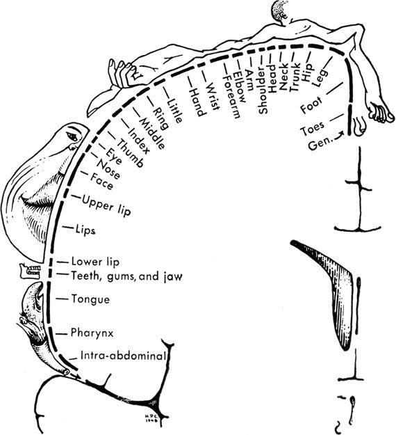
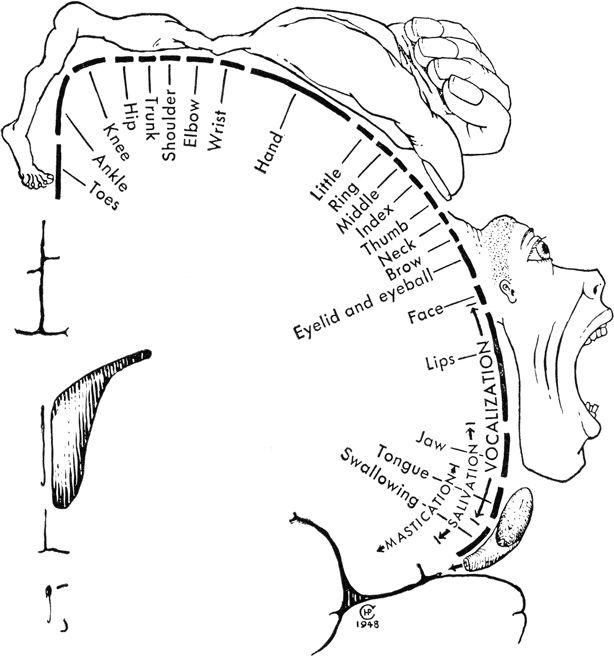
Later studies by the American-born neurosurgeon Wilder Penfield in Montreal observed the response of conscious epilepsy patients to brain stimulation by electrodes. Penfield used the technique in order to plan brain surgery to relieve convulsions experienced in specific regions of the body. But what he obtained as a result was a new map of the brain. Penfield’s masterstroke was to employ an artist when he published his findings in 1937. Mrs H. P. Cantlie drew a ‘cortical homunculus’ in which the various sensory and motor functions of the body were depicted at a scale in proportion to the volume of the area of the brain thought to be responsible for their control. Unfortunately, the diagram – showing greatly enlarged thumbs and large fingers, hands and feet compared to the limbs and trunk of the body – looked a bit like a frog squashed on the road. More instructive and enduring is a later version that Penfield published in which the sensory and motor organs are draped directly around the hemispheres of the brain in a sectional view across the head. The lips and the thumb stand out especially. This graphic idea has taken root and inspired ever more grotesque variants since, as well as having its precursors in the homunculus of medieval belief, who was literally a little man, or ‘manikin’, a kind of Mini-Me that might be conjured by an alchemist or a magician. These distorted human figures are perhaps also imaginatively invoked in the gangly dragons and monsters of our nightmares and cartoons, with their grasping fingers and clumping feet.

True images of the brain are brought to us by a different magic. The secret is the phenomenon of nuclear magnetic resonance, a discovery of such momentous significance that it has been marked by the award of Nobel Prizes on six occasions: three in physics, two in chemistry, and the latest in medicine, awarded in 2003 for its application in the form of medical imaging now universally known as MRI.
I went to have my brain scanned more than twenty years ago. It was the spring of 1988, and this form of imaging had only just gained approval for clinical use. So new was the technique that nobody had yet thought to ditch the ‘nuclear’ part of the name that somehow failed to reassure prospective patients. I am not a patient, however, but writing an article for
Popular Science
magazine.
When I arrive at the Albany Medical Center in the New York state capital, the white-coated head of neuroradiology, Gary Wood, begins by asking me some preliminary questions. ‘Is there anything wrong with you? Do you have anything metal on you – pens, paper clips?’ I deposit keys, a pen, and my tape recorder in a locker. Then the doctor opens a big door shielded with copper and ushers me into the MRI room.

A large doughnut-shaped machine fills the room. It is emblazoned with the logo of General Electric, the company founded nearly 100 years before by Thomas Edison, funnily enough, and based in nearby Schenectady. Smoothly contoured white plastic conceals its five-tonne magnet. (Medical NMR magnets may generate magnetic fields measuring some 15,000 gauss; the Earth’s magnetic field by comparison averages just 0.5 gauss, while the magnet in your fridge door might produce around 50 gauss.) Wood’s assistant helps me on to a gurney that projects from the bore of the magnet, and then flicks a switch. Powered by hydraulics (motors won’t work near this huge magnet), I glide almost silently into the magnet until my head is positioned at its centre. Any sense of claustrophobia is mitigated by the mirror thoughtfully angled above my eyes so that I can see out beyond my feet and through the room’s observation window to where Gary and his colleagues are monitoring the scan. Through a two-way audio link, I hear them typing instructions into the computer and chatting excitedly about their new equipment. ‘Lie still,’ I am told. Gary presses a button. A rapid, dull drumming fills my ears, but I feel nothing as the massive machine scans the depths of my brain.
Afterwards, Gary shows me what he has recorded on the monitor. It is the first time I have been able to see inside my own body. Yet even at this early date, I find I am jaded by the generic familiarity of the images. ‘MRI has shifted our sense of transparency so that we can see those structures whose form and function had previously been the domain of poets and philosophers,’ I read in one rather awestruck history of medical imaging. But what is seeing? I am aware that what I am looking at is not a simple photograph, but a highly indirect image, a digital manifestation of a series of radio-frequency signals, which are themselves the product of tiny magnetic fields produced by hydrogen atoms in my brain in response to the massive input signal of the imaging machine. It seems to me the poets and philosophers might still have the edge.
Sensing my ambivalence, perhaps, Gary points to different shades of grey on the screen that represent the outer shell of my skull, my bone marrow, and even my cerebrospinal fluid. ‘Now we’re going to page through,’ he tells me. ‘We’re going to drive right through your head.’ A series of images appears on the screen as Gary chases my optic nerves from my eyes into my brain. He pauses at one picture, a cross-section clearly showing my nose, throat and sinuses. ‘Here’s something that looks like a Dristan commercial,’ he laughs. As I depart, he gives me a souvenir print of my scan. Sadly, I no longer have the image, so I cannot tell whether my parietal lobe is expanded or my lateral sulcus closed up like Einstein’s.
Improvements in magnetic resonance imaging made since the time of my scan now allow scientists to obtain live, moving images of the working brain. Experiments in
functional
magnetic resonance imaging (fMRI) typically involve scanning a subject’s brain while he or she performs particular tasks. This yields images that highlight the parts of the brain that are temporarily more active. The digital image processing applied to the MRI scans generally displays a section through the whole brain in black-and-white with the active area shown as a coloured highlight. Thanks to this manipulation, we now speak happily of the parts of the brain that ‘light up’ when we think particular thoughts, although, strictly speaking, the observed ‘lighting up’ is an indication of increased blood flow and not necessarily of a particular mental activity.
This new technology is an important aid in diagnosing brain disease, but it also provides a new tool for investigating the way the brain works normally. Many studies are underway to examine aspects of human mental activity that we tend to regard as important in defining who we are as individuals. These include the making of moral choices, the display of prejudice, and the exercise of personal creativity. Even simple decisions without consequences require the exercise of choice, which is an expression of personality. Neuroscientists at the Oxford Centre for Functional MRI of the Brain devised an experiment that required subjects to push buttons in order to switch from an arbitrary state A to states B or C. When subjects chose freely, their action was accompanied by increased activity in one particular part of the brain and reduced activity in another. When the same subject was directed as to what to do by a second person, however, this picture was reversed. The experiment seems to show that the neural mechanisms underlying our assessment of the choices we make are different according to whether those choices are forced or freely made.
But what about a real moral dilemma? Joshua Greene at Harvard University asked his subjects to imagine a situation in which a crying baby threatens to give away the presence of a group of people hiding from enemy soldiers: do you smother the baby to save the lives of the others? His results showed that brain regions associated with planning, reasoning and attention were comparatively more active when people chose to harm some individuals in order to save others. In other words, people think harder when what they decide will have consequences for others. It is what we would at least hope for of our fellow human beings.
Greene’s colleague at Harvard, Jason Mitchell, has been using fMRI to investigate empathy and prejudice. Understanding other people involves imagining ourselves in their position. This is easier to do when the other person is similar to oneself. Mitchell asked his subjects, defined according to their social and political beliefs, to evaluate imagined persons both strongly like and strongly unlike themselves. The brain images he recorded show that the perception of a similar ‘other’ engages a region of the brain known to be linked to self-referential thought, whereas perception of a dissimilar ‘other’ activates a different region. It does not reveal
why
, but it does show a little of what happens
when
, for example, white people more readily associate black faces with negative attributes and white faces like their own with positive ones. Such work may provide a key to understanding racial and other forms of prejudice.
Creative works such as paintings, symphonies and novels are seen as highly personally expressive. But can the creative process be seen as it happens in the brain? Charles Limb at the Johns Hopkins School of Medicine in Washington, DC has tried to catch a glimpse of it by recording fMRI scans of skilled jazz musicians as they improvise at the piano – devising music never thought of or played before. An average made of the brain images of a number of improvisers shows particular areas of the brain activated and others deactivated, suggesting that creativity, too, is localized. Imaging studies of the normal brain such as these gain validity by taking data from a sample population of subjects, not just a single person. I can see how it might be dangerous to interpret one person’s scan in a particular way when looking at something as personal and subjective as prejudice or creativity. Yet I can’t help wondering if these statistical aggregates, like Galton’s composite photographs, risk throwing away the very information they are trying to gather.
