Authors: Sebastian Seung
Connectome (22 page)

To find connectomes, we will have to create machines that produce clear images of neurons and synapses over a large field of view. This will be an important new chapter in the history of neuroscience, which is perhaps best seen not as a series of great ideas but as a series of great inventions, each of which surmounted a once insuperable barrier to observing the brain. It now seems trivial to say that the brain is made of neurons, but the path to this idea was tortuous. And for a simple reasonâfor a long time, it was impossible to see neurons.
Â
Living sperm were first observed in 1677 by Antonie van Leeuwenhoek, a Dutch textile merchant turned scientist. Leeuwenhoek made the discovery with his homebrew microscope, but he didn't fully recognize its significance.
He did not prove that the sperm, rather than the surrounding fluid in semen, were the agents of reproduction. And he had no inkling of the process of fertilization by which an egg and a sperm unite. But by paving the way to these discoveries by his successors, Leeuwenhoek's work was epoch-making.
Three years earlier, Leeuwenhoek had examined a droplet of lake water with his microscope. He saw tiny objects moving around and decided that they were alive. He called them “animalcules”
and wrote a letter about them to the Royal Society of London. By now we are completely used to the idea of microscopic organisms and have difficulty imagining how much they must have stunned his contemporaries. At the time, though, Leeuwenhoek's claims seemed so fantastic that they provoked suspicions of fraud. To allay these fears, he sent the Royal Society testimonial letters from eight eyewitnesses, including three clergymen, a lawyer, and a physician.
After several years his claims were finally vindicated, and the Society honored him with membership.
Leeuwenhoek is sometimes called the father of microbiology. This field turned out to have great practical significance in the nineteenth century, when scientists like Louis Pasteur and Robert Koch showed that microbial infection can cause disease. Microbiology was also critical in the development of the cell theory. This cornerstone of modern biology, formulated in the nineteenth century, holds that all organisms are composed of cells. Microscopic organisms are those that are composed of just a single cell.
Most of the members of the Royal Society were wealthy men with the time to devote themselves to intellectual pursuits. Leeuwenhoek was not born rich, but by age forty he had secured enough income to turn his attention to science. He did not study at a university, and did not know Latin or Greek. How did this self-educated man from humble origins achieve so much?
Leeuwenhoek did not invent the microscope; the credit goes to eyeglass craftsmen who worked at the end of the sixteenth century. Like today's microscopes, the first ones combined multiple lenses, but they could magnify only 20 to 50 times. Leeuwenhoek's microscopes delivered up to 10 times better magnification with a single, very powerful lens.
We can't be certain how he learned to make such outstanding lenses, because he kept his methods secret. This was Leeuwenhoek's “unfair” advantage: He made better microscopes than the ones used by his rivals.
When Leeuwenhoek died, his methods were lost. Later, in the eighteenth century, technical improvements made the multilens (“compound”) microscope more powerful than Leeuwenhoek's. Scientists were able to see the structures of plant and animal tissues more clearly, which resulted in the acceptance of the cell theory in the nineteenth century. Yet there was one place where the theory ran into trouble: the brain. Microscopists could see the cell bodies of neurons and the branches extending from them. But they lost track of the branches after a short distance. All they could see was a dense tangle, and no one knew what happened there.
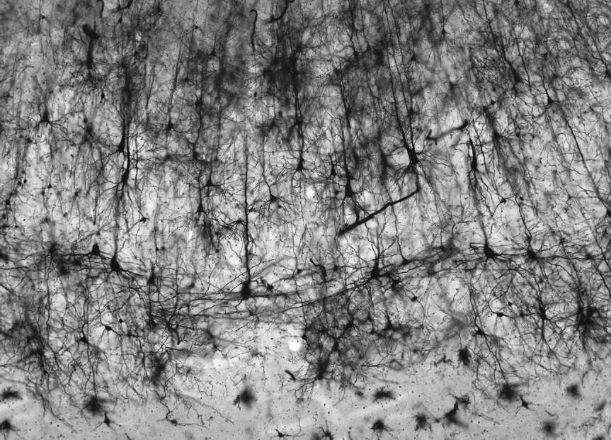
The problem was resolved by a breakthrough in the second half of the nineteenth century. An Italian physician named Camillo Golgi invented a special method of staining brain tissue. Golgi's method stains only a few neurons; it leaves almost all of them unstained and hence invisible. Figure 26 may still look a little crowded, but we can make out the shapes of individual neurons.
Golgi's scientific rival, the Spanish neuroanatomist Santiago Ramón y Cajal, was presumably viewing something like this in his microscope when he drew the image shown in Figure 1.
Â
 Â
Â
Â
Figure 26. Golgi staining of neurons in the cortex of a monkey
Â
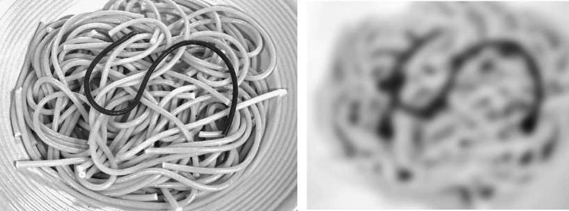
Golgi's new method was an extraordinary advance. To appreciate why, let's imagine the branches of neurons as entangled strands of yellow spaghetti. (I introduced this metaphor earlier, but it's now even more apt, given Golgi's national origin.) Cooks with extremely bad eyesight see only a yellow mass on the plate, because the individual strands are too blurry to be distinguished. Now suppose that a single dark strand
is mixed in with the others (see Figure 27, left). Even with blurry vision, it's possible to follow the path of the dark strand (right).
Â
 Â
Â
Â
Figure 27. Why Golgi staining works: photograph of pasta before
(left)
and after
(right)
blurring
Â
As an invention, a microscope might seem more glamorous than a stain. Its metal and glass parts are impressive, and can be designed using the laws of optics. A stain isn't much to look at; it might even smell bad. Stains are often discovered by chance rather than design. Actually, we still don't know why Golgi's stain
marks only a small fraction of neurons. All we know is that it works. In any case, Golgi's stain and others have played an important role in the history of neuroscience. “The gain in the brain lies mainly in the stain,” neuroanatomists like to say. Golgi's is simply the most famous.
Science can languish for a long time if the proper technology does not exist. Without the right kind of data, it can be impossible to make progress, no matter how many smart people are working on the problem. The nineteenth-century struggle to see neurons lasted until the invention of Golgi's staining method, which was soon used most avidly, and most illustriously, by Cajal. In 1906 Golgi and Cajal voyaged to Stockholm to receive the Nobel Prize “in recognition of their work on the structure of the nervous system.” As is customary, both scientists gave special lectures describing their research. But rather than celebrate their joint honor, the two men took the opportunity to attack each other.
They had long been embroiled in a bitter dispute. Golgi's staining method had finally revealed neurons to the world, but the limited resolution of the microscope still left ambiguities. When Cajal looked in his microscope, he saw points at which two stained neurons contacted each other but still remained separate. When Golgi looked in
his
microscope,
he saw such points as locations where neurons had fused together into a continuous network, forming a kind of supercell.
By 1906 Cajal had convinced many of his contemporaries that a gap existed, but it was still unclear how neurons could communicate with each other if they were not physically continuous. Three decades later, Otto Loewi and Sir Henry Dale were awarded the Nobel Prize “for their discoveries relating to chemical transmission of nerve impulses.” They had found conclusive evidence that neurons can send messages by secreting neurotransmitter molecules, and receive messages by sensing them. The idea of a chemical synapse explained how two neurons could communicate across a narrow gap.
Still, no one had actually seen a synapse. In 1933 the German physicist Ernst Ruska built the first electron microscope, which used electrons rather than light to yield much sharper images. Ruska moved to Siemens and developed a commercial product. After World War II the electron microscope grew in popularity. Biologists learned how to cut their specimens into extremely thin slices and then image the slices. Finally, they had clear pictures.
The first images of synapses, obtained in the 1950s, showed that two neurons did not fuse at a synapse. There was definitely a boundary separating the two cells, and sometimes one could even see an extremely narrow gap between them. These are features that cannot be seen clearly with a light microscope, which is why Golgi and Cajal had been unable to resolve their debate.
With this new information, victory to Cajal. Or so it seemed. In the end, Golgi turned out to be right as well. In addition to chemical synapses, the brain also contains electrical synapses, as I mentioned earlier. At this type of synapse, special ion channels span the cleft between the two membranes and function as tunnels through which ions, electrically charged atoms, can travel from the inside of one neuron to the inside of another. An electrical synapse carries electrical signals directly between neurons without the need for an intermediate chemical signal, and effectively fuses two cells into one continuous supercell, as Golgi envisioned.
I've billed the electron microscope as the invention that enabled the imaging of synapses. But new stains
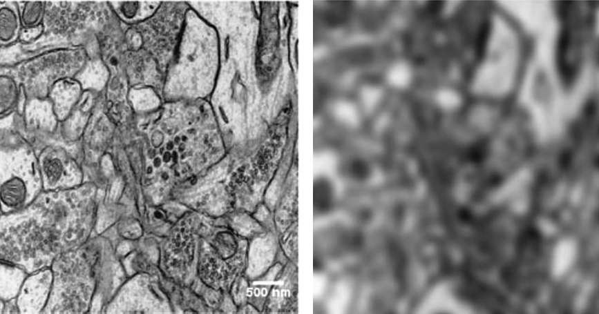
were also crucial. With electron microscopy, it's sensible to use “dense” staining methods, which mark all neurons. The combination of electron microscope and dense stain revealed what neuroscientists had already imagined but had never seen clearlyâthe entangling of the branches of many neurons. While the Golgi stain showed the shape of a neuron, it gave the false impression that neurons are islands surrounded by large expanses of nothing. In fact, brain tissue is packed full of neurons and their branches, as you can see at the left in Figure 28.
This image resembles what you'd see if you cut through a tangled mass of spaghetti. The cut ends of the individual strands would have circular or elliptical cross-sections like the cross-sections of neural branches in the image.
The laws of physics limit the resolving power of a light microscope to the wavelength of light, which is a fraction of a micrometer. Details smaller than this are blurred, a barrier known as the diffraction limit.
Shown at the right in Figure 28 is another version of the electron microscope image, this one artificially blurred to simulate how it would look in a light microscope.
The cross-sections of the thinnest branches of neurons are no longer clearly visible. That's why sparse methods of staining like Golgi's, which mark only a few neurons, were necessary when using the light microscope. The electron microscope's much higher resolving power makes it possible to see all neurons at the same time with dense staining.
Â
 Â
Â
Â
Figure 28. Cross-sections of axons and dendrites imaged by an electron microscope, before
(left)
and after
(right)
blurring
Â
An electron microscopic image shows only two-dimensional cross-sections of neurons, however. To see neurons in their full glory, we need three-dimensional images. These can be obtained by slicing up brain tissue using a high-tech version of the machine in your local delicatessen, and then imaging every slice. Cutting might sound trivial, but the slices must be tens of thousands of times thinner than your typical prosciutto. For this, we need a most unusual knife.
