Pediatric Examination and Board Review (246 page)
Read Pediatric Examination and Board Review Online
Authors: Robert Daum,Jason Canel

(B) arthritis, especially of the ankles and knees, is common
(C) in a small percentage of children, the purpuric rash is secondary to thrombocytopenia
(D) edema of the scalp, hands, and feet is not uncommon, especially in affected children younger than 4 years old
(E) skin biopsy will show a leukocytoclastic vasculitis with immune globulin (Ig)A deposition
18.
All the following are true about GI involvement in HSP except
(A) GI hemorrhage is common and may be occult or gross, presenting as melena
(B) GI symptoms can present before the rash
(C) a normal barium enema rules out intussusception in HSP
(D) complications include bowel wall infarction and perforation
(E) ultrasonography may show edema of bowel wall and may identify an intussusception
19.
Which statement is true for renal involvement secondary to HSP?
(A) most children who have hematuria during the acute phase of HSP illness have progression of renal disease
(B) it is associated with a membranous lesion on renal biopsy
(C) it usually presents shortly after HSP diagnosis with nephrotic syndrome and hypertension
(D) it may persist in 1-5% of children and may progress to end-stage disease in approximately 1%
(E) it occurs more commonly in patients younger than 8 years old at the time of HSP diagnosis
20.
An 18-month-old boy was healthy until the acute onset of persistent fevers 4 days ago. He has been irritable but alert. His mother brings him to the emergency department and you note an exanthem on the trunk. You suspect Kawasaki disease (KD). You may expect to see all but which of the following findings on your admission physical?
(A) desquamation of the skin on the palms and soles
(B) cervical adenopathy
(C) strawberry tongue and dry, cracked lips
(D) edema on the dorsum of hands and feet
(E) conjunctivitis
21.
A 15-year-old girl has had Raynaud symptoms for 1 year, and she occasionally develops sores on her fingertips. Previous laboratory assessment by her pediatrician revealed a normal CBC, C3, C4, and UA but a positive ANA (1:2560 nucleolar pattern). Over the past 6 months, she developed fullness of her fingers with skin tightening and decreased finger-joint range of motion. A diagnosis of systemic sclerosis (scleroderma) is suspected. Further workup should include
(A) renal biopsy
(B) brain magnetic resonance imaging (MRI)
(C) muscle biopsy
(D) 24-hour urine collection for protein and creatinine clearance
(E) pulmonary function tests
22.
In the past 6-12 months, an 8-year-old boy developed an altered appearance of his lower right leg including tautness and shininess of the skin and decreased ankle range of motion. He was diagnosed by the rheumatologist to have linear scleroderma. Which of the following physical findings does not occur in this entity?
(A) muscle atrophy
(B) Raynaud
(C) shortening of the involved limb
(D) occasional patches of morphea at distant locations
(E) flexion contractures on the involved limb
Match each entity in questions 23 through 33 to one descriptive characteristic, A-K.
23. | (A) large vessel vasculitis |
24. | (B) + cANCA (antineutrophil cytoplasmic antibodies) |
25. | (C) severe uveitis of anterior and posterior uveal tracts |
26. | (D) a type of panniculitis granulomatosis |
27. | (E) sicca complaints (dry eyes and ↓ oral secretions) |
28. | (F) vasculitis of small and medium-sized vessels |
29. | (G) presents 7-14 days after antigen exposure |
30. | (H) elevated anti-DNase B titers |
31. | (I) linear scleroderma of the face |
32. | (J) noncaseating granulomas |
33. | (K) treatment with intravenous gammaglobulin (IVIG) |
ANSWERS
1.
(E)
The patient’s clinical complaints, physical findings, and CBC results are all nonspecific and are not diagnostic for any particular disease. It is important to have a broad differential when first assessing this type of patient. Malignancy, such as leukemia, should be considered in children with this clinical history and depressed cell lines on CBC. Children with leukemia may present with musculoskeletal pain that is frequently out of proportion to the physical findings. This diagnosis may be overlooked in those who do not have blasts on peripheral smear (approximately 15% of children). SLE should be considered in any patient who presents with multisystem complaints. Constitutional symptoms such as fever, fatigue, and weight loss are common in SLE, as is joint involvement, which is present in approximately 75% of patients. Mild cytopenias are common laboratory findings in SLE. EBV infection, especially in the teenager, can present with multisystem complaints similar to those seen in SLE, including constitutional symptoms, arthralgias/ arthritis, and adenopathy. Children with EBV infections often have cytopenias and positive ANAs, which can add to the challenge of differentiating between SLE and EBV. Further laboratory evaluation (EBV antibody profile, complement levels, autoantibodies such as anti-Sm and anti-double stranded DNA, UA) may help guide the clinician to the correct diagnosis.
2.
(E)
ANAs are found in many clinical settings, even among healthy individuals (especially among those who have primary relatives with autoimmune disease or in the elderly). They may be detected in infections (eg, EBV, streptococcal, subacute endocarditis, hepatitis C), autoimmune disease (SLE, mixed connective tissue disease, dermatomyositis, scleroderma, juvenile idiopathic arthritis, adult rheumatoid arthritis, Sjögren syndrome), druginduced processes (eg, secondary to anticonvulsants, isoniazid, penicillamine, hydralazine, procainamide, minocycline), and organ-specific autoimmune disease (autoimmune hepatitis, thyroiditis). ANA is present in almost all patients with SLE (>98%). Titers are variable in SLE but are usually 1:320 or higher. It is important to recognize that although virtually all patients with SLE have a positive ANA, most people with a positive ANA do not have SLE.
3.
(B)
Anti-dsDNA and anti-Sm are specific for SLE and are present in approximately 70% and 20% of SLE patients, respectively. Other autoantibody subtypes may be present but are not specific for SLE, and they may be identified with other autoimmune entities:
| SPECIFIC AUTOANTIBODY | CLINICAL ASSOCIATIONS |
| Anti-dsDNA | SLE |
| Anti-Sm | SLE |
| Anti-U1-RNP | Mixed connective tissue disease; SLE |
| Anti-Ro/SS-A | SLE; Sjögren syndrome; neonatal lupus; C2 and C4 deficiencies |
| Anti-La/SS-B | Sjögren syndrome; SLE; neonatal lupus |
| Anti-Scl 70 | Systemic sclerosis |
| Anti-Jo-1 | Polymyositis, especially with interstitial lung disease |
| Antihistone | Drug-induced lupus; SLE |
| Anticentromere | Limited scleroderma (CREST) |
Abbreviation: CREST, calcinosis cutis, Raynaud phenomenon, esophageal motility disorder, sclerodactyly, telangiectasia.
4.
(A)
The American College of Rheumatology 1997 criteria for classification of SLE are helpful diagnostic tools for SLE diagnosis. The criteria are
• Malar rash (
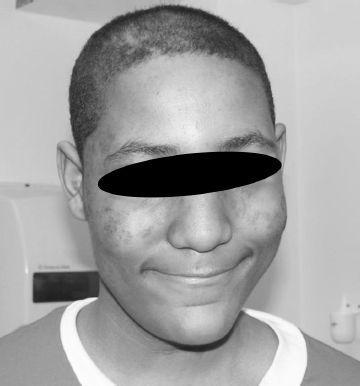
Figure 141-1
)
• Discoid rash
• Photosensitivity
• Oral or nasal ulcerations
• Nonerosive arthritis
• Polyserositis
• Nephritis (proteinuria >0.5 g/day or cellular casts)
• Encephalopathy (seizures or psychosis)
• Cytopenia
• Positive ANA
• Positive immunoserology: +anti-dsDNA or +anti-Sm, or presence of antiphospholipid antibodies

FIGURE 141-1.
Malar rash in a teenage boy with systemic lupus erythematosus. See color plates.
The presence of 4 of these 11 criteria has a sensitivity and a specificity of more than 95% for the diagnosis of SLE. SLE should be considered in children who present with multisystem complaints and signs. Although not included in the criteria just listed, several other features may be present that increase suspicion for SLE, such as constitutional symptoms (fatigue, weight loss, fevers), alopecia, myalgias/myositis, Raynaud phenomenon, lymphadenopathy, hepatosplenomegaly, myocarditis, and thrombosis. Decreased C3 and C4 levels in a patient with a positive ANA associated with any of the symptoms just listed raise concern of a diagnosis of SLE. In 1999, the American College of Rheumatology (ACR) developed expanded nomenclature for neuropsychiatric SLE when it described 19 central and peripheral neurologic syndromes, including cognitive dysfunction, headaches, and cranial and polyneuropathies. Answer A, erythema marginatum, is a skin finding in acute rheumatic fever.
5.
(D)
The arthritis in SLE patients is rarely erosive, and joint deformity is uncommon. A small percentage of patients with SLE may develop Jaccoud arthropathy, which is a nonerosive but deforming arthropathy.
6.
(C)
Lupus nephritis occurs in more than 60% of children with SLE. The risk of renal involvement is increased among those with antibodies to dsDNA but not among those with anti-Sm antibodies. Although some patients may present with nephrotic syndrome, hypertension, and renal insufficiency, the majority initially have no clinical symptoms of renal disease. Therefore, it is important to perform frequent urinalyses, assess protein excretion (the easiest way is by performing a protein-to-creatinine ratio on a spot urine), and follow the serum creatinine and albumin. Rarely, patients with a normal UA may have renal involvement. Because urine and blood studies are indirect assessments of renal status in SLE patients, a kidney biopsy is often necessary to provide important information to help with treatment decisions. The 2004 International Society of Nephrology (ISN) classification system, which replaced the World Health Organization (WHO) classification system, categorizes renal lesions based on light (and occasionally electron) microscopy, and immunofluorescence. The ISN classification describes 6 classes, which are then further subdivided to provide more detail of the renal pathology:
