Pediatric Examination and Board Review (40 page)
Read Pediatric Examination and Board Review Online
Authors: Robert Daum,Jason Canel

(E) skin tags
7.
The patient’s mother reports that her mother had similar skin changes and died of a tumor. The most likely malignancy she had was
(A) malignant neurofibroma
(B) basal cell carcinoma
(C) melanoma
(D) leiomyosarcoma
(E) malignant plexiform neurofibroma
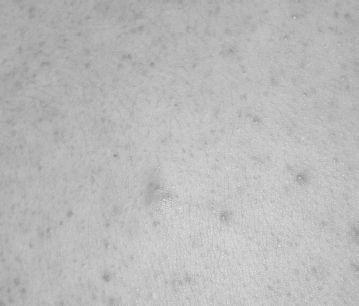
FIGURE 21-2.
See color plates.
8.
Another 6-month-old Hispanic boy presents for well-child care. His parents note that they have become increasingly aware of light patches of skin. They are unsure whether they were present at birth because the child had much fairer skin at that time. Examination reveals six hypopigmented oval patches on the trunk and extremities measuring 1-5 cm in diameter (
Figure 21-3
). The presence of three hypopigmented macules in infancy is
(A) a diagnostic criterion of tuberous sclerosis
(B) common in the general population
(C) a risk factor for the development of vitiligo
(D) typical of Klippel-Trenaunay syndrome
(E) a marker of photosensitivity
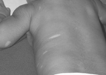
FIGURE 21-3.
See color plates.
9.
The mutated gene associated with tuberous sclerosus is
(A) neurofibromin
(B) hamartin
(C) fibrillin
(D) profilaggrin
(E) retinoblastoma 1 (RB1)
10.
The following skin lesion of tuberous sclerosus is usually present at birth
(A) angiofibroma
(B) Shagreen patch
(C) confetti macules
(D) periungual fibroma
(E) none of the above
11.
The following lesion is considered pathognomonic of tuberous sclerosus
(A) periungual fibroma
(B) forehead fibrous plaque
(C) molluscum fibrosum pendulum
(D) Shagreen patch
(E) confetti spots
12.
The most common early seizure type observed in tuberous sclerosis is
(A) partial
(B) infantile spasm
(C) absence
(D) febrile
(E) tonic-clonic
13.
The mutated gene of tuberous sclerosus normally functions as
(A) a tumor suppressor
(B) epidermal growth factor
(C) gap junction protein
(D) cell cycle inhibitor
(E) apoptosis initiator
14.
The cardiac lesion most closely associated with tuberous sclerosus is
(A) coarctation of the aorta
(B) patent ductus arteriosus
(C) rhabdomyoma
(D) congenital heart block
(E) tetralogy of Fallot
15.
An 11-year-old boy with reddish shiny papules of the face, seizures, mental retardation, and newly noted hypertension should be evaluated for
(A) pheochromocytoma
(B) renal artery stenosis
(C) hyperthyroidism
(D) renal cyst
(E) diabetes mellitus
16.
Cortical tubers associated with tuberous sclerosis
(A) are lesions of bone
(B) are seen on X-ray
(C) are astrocytomas
(D) have malignant potential
(E) may correlate with the occurrence of seizures and mental retardation
17.
A 9-month-old girl presents for routine well-child care and you note a red purple patch on the left forehead, extending onto the upper eyelid, and posteriorly into the hairline (
Figure 21-4
). This lesion most likely represents a
(A) hemangioma
(B) port wine stain
(C) café-au-lait macule
(D) congenital nevus
(E) salmon patch
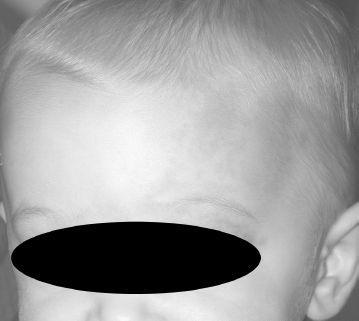
FIGURE 21-4.
See color plates.
18.
The patient is noted to have delayed developmental milestones. You inform the parents that further evaluation should include a
(A) head MRI
(B) head ultrasound
(C) skin biopsy
(D) echocardiogram
(E) karyotype
19.
Additionally, it is important that the patient be evaluated by a
(A) pediatric orthopedist
(B) pediatric cardiologist
(C) pediatric ophthalmologist
(D) geneticist
(E) pediatric gastroenterologist
ANSWERS
1.
(B)
The most common diagnosis in the setting of multiple café-au-lait macules is neurofibromatosis. Optic gliomas peak in incidence between 4 and 6 years of age in patients with neurofibromatosis type I. The description of tan-brown well-demarcated patches is most suggestive of café-au-lait macules and not congenital nevi because nevi are usually more irregularly colored and often have a raised or infiltrative component. Retinal phakomas and periungual fibromas are associated with tuberous sclerosus.
2.
(D)
Multiple café-au-lait macules may be seen in numerous conditions and sometimes in isolation without other findings. McCune-Albright, Bloom, and Turner syndromes may all demonstrate multiple café-au-lait macules. The diagnostic criteria for neurofibromatosis state that the café-au-lait macules must be greater than 5 mm in diameter in children (>15 mm after puberty) and greater than 5 in number. Sturge-Weber syndrome involves a facial port wine stain and intracranial vascular malformations; café-au-lait macules are not seen more frequently in this condition.
3.
(A)
Axillary and inguinal freckling (Crowe sign) is another diagnostic criterion for neurofibromatosis type I. These findings may not appear until 3-5 years of age. A Shagreen patch, ash leaf spot, and periungual fibromas are seen in tuberous sclerosis. Mucosal lentigines are features of Peutz-Jeghers syndrome.
4.
(C)
A first-degree relative with findings consistent with neurofibromatosis can confirm the diagnosis when only multiple café-au-lait macules are present at the time of evaluation. Neurofibromatosis may run a benign course and individuals may not be aware of their diagnosis; therefore, a careful examination of first-degree relatives can be a very important diagnostic procedure.
5.
(A)
A slit-lamp exam can facilitate visualization of Lisch nodules on the iris, which constitutes another diagnostic criterion to aid in the diagnosis of neurofibromatosis. Lisch nodules may not be present in infancy, however, and therefore a negative exam will not rule out the diagnosis.
6.
(D)
Neurofibromas develop in individuals with neurofibromatosis over time. The presence of two or more satisfies one of the diagnostic criteria. These growths are typically soft and spongy and may be flesh colored to tan-brown or violaceous in color. They are typically asymptomatic, although sometimes patients report tenderness. Neurofibromas represent growth of nerve sheaths.
7.
(E)
The presence of one plexiform neurofibroma is a diagnostic criteria of neurofibromatosis type I. Plexiform neurofibromas are growths of a nerve sheath that extend along the length of a nerve and include multiple fascicles. These lesions may be located superficially in the dermis or more deeply in the soft tissue. There may be associated soft tissue and bony overgrowth. Malignant change may occur within these tumors. Some plexiform neurofibromas are noted to have an overlying giant café-au-lait macule, and there seems to be a higher incidence of malignant change within these lesions. Basal cell carcinomas are the most common skin cancer but are rarely fatal.
8.
(B)
Hypopigmented macules are common in the general population, with a prevalence as high as 4.7% in a white population. Higher incidences are typically reported in darker-skinned populations. There are not strict diagnostic criteria regarding skin lesions in tuberous sclerosus. It is generally stated that up to three hypopigmented macules in an otherwise healthy individual without a family history of tuberous sclerosus should not necessitate further workup.
9.
(B)
Hamartin and tuberin are tumor suppressor genes associated with tuberous sclerosus. Neurofibromin is the mutated gene of neurofibromatosis type I. Fibrillin is the mutated gene of Marfan syndrome. Profilaggrin is the mutated gene of ichthyosis vulgaris.