The Psychopath Inside (5 page)
Read The Psychopath Inside Online
Authors: James Fallon

As I transitioned into the upperclassman years, I renewed my interest more and more in the biological and chemical sciences, and became an even stauncher believer in the notion that behavior is all about chemistry and electricity and probably genes, and that if one could manipulate these genetic processes, one could control the brain and the mind. The film
Charly
, based on the novel
Flowers for Algernon,
came out in 1968, while I was a third-year biology
student. It highlighted the biochemical basis of behaviors and resonated with me at a time when I was really receptive to it. My career as a mechanistic, reductionist, genes-control-all scientist had begun. The rest, a belief in free will and God, went missing that junior year.
Around that time, I, the former Catholic Boy of the Year in my diocese of New York, left the Catholic Church. I approached one of my professors, Father Stapleton, and told him about my doubts and asked for a formal last confession. He laughed and said, “We don't usually help people get out of the Church,” but he agreed. I was still well behaved, and studious of the Scripture. I'd thoroughly learned the lessons of Christ and Aquinas and Augustine. He said, “You don't need the Church anymore, and in fact it's making you crazy, with all the OCD stuff.” With that, a great dismal onus lifted, and I felt free and light. It was like a switch had flipped in my brain, one full of positive and aggressive energy, buoyed by self-confidenceâmaybe even overconfidence.
My belief that we are born and not made also had a profound impact on my political views. Whereas prior to college I subscribed to a mix of my mother's conservatism and my aunts' liberalism (my father was neutral), I became increasingly fed up with views on both the left and the right that environmental forces are somehow responsible for shaping who we are. For the right, this manifested itself in support of the nuclear, heterosexual family; on the left, it was rooted in the belief that society should take care of its citizens. In 1969, I became a Libertarian.
The possibilities available in a career in the neurosciences,
where one deals in hard science and facts, were intoxicating to me, and I would commit my life to the study of how the brain shapes who we are. Soft psychology, although of interest to me throughout high school and the early college years, seemed to offer few real insights into what makes us human. After some temporary academic flubs and fumbles in my senior year of college, I would first teach in an all-girls Catholic high school in Albany, and then enter the physiological psychology and psychophysics graduate program at Rensselaer Polytechnic Institute in Troy. After that, I entered a doctoral program in anatomy and physiology at the University of Illinois College of Medicine in Chicago, studying, curiously enough in hindsight, the orbital cortex and temporal lobe and associated systems in the primate brainâareas I later saw damaged in killers' brains. This positioned me on a bright-line trajectory to a neurochemistry and neuroanatomy postdoctoral stint at the University of California, San Diego, before landing me a tenure-track job at the University of California, Irvine, where I have been ensconced in the ensuing years, as a satisfactorily successful professor to this day. All had been beautiful and terrifically fulfilling and easy the whole way through from college onward.
Smooth, at least, for thirty-five years.
The Brain of a Killer
I
first became interested in science when I was a child, thanks to my early experiences working on a farm, walking in the forest, and investigating life in ponds and streams in upstate New York. I was urged on in my interest of the world of bugs, frogs, and creepy crawlies by my parents and grandparents, and especially my aunt Flo, who was a nurse and graduate of Columbia University. Flo saw my interest in the natural world starting in the first years of grade school. I asked her once when she first had that insight, and she said that when I was nine months old she was bathing me in the kitchen sink, and when she drained the large porcelain basin, I had a wide-mouthed, gaping look of amazement as the water swirled down the drain. From that moment onward, she said, I was a scientist.
When we moved from Cohoes to Loudonville in 1956, Aunt Flo gave me a microbiology text from her nursing school class at Columbia, at about the same time my father gave me an old, but high-quality, vintage 1930s Bausch & Lomb microscope. I was in the fourth grade.
Oddly enough, at the same time that I developed a fascination with science and nature, I harbored my growing obsession with
religion and spirituality. I began to ponder the infinite and the hereafter. Whatever had put these worries into my head, the combination of awe and fear they inspired was both thrilling and terrifying and led to a lifelong quest to understand the fundamentals of the human mind, heart, and soul.
For the first twenty years of my academic life I devoted all my work to the basic neurosciences, while also teaching medical students and graduate students about the structure and function of all the systems in the body in the gross anatomy and microanatomy courses. In the 1990s, I started teaching more and more in UCI's human neurosciences curriculum to medical students, graduate students, and residents in neurology and psychiatry, and this whetted my appetite for understanding the biological basis of the human mind, both normal and abnormal. As I became more and more knowledgeable about the human brain, as opposed to just animal brain neuroanatomy, I was being asked by more and more colleagues in psychiatry and in the behavioral and cognitive sciences to analyze brain scans of their patients in the clinical trials they were conducting for drug companies. I was developing a reputation for knowing about the entire brain and nervous system, and this lack of specialization fit my childhood dream of becoming a Renaissance man, like my hero, Leonardo da Vinci. So instead of becoming an expert in something, I was actually becoming an expert in nothing at all.
One day in 1995, my colleague in psychiatry Anthony rang me up and said, “Hey, Jim, I've got a job for you. These lawyers I'm consulting have a guy who murdered some people, and we did
a scan to see if there was something wrong with his brain. Could you take a look and tell us what you see?” I said sure and reviewed his PET scan.
A PET (positron emission tomography) scan is a tool used in radiology to determine the functioning of the body, specifically small areas the size of a grain of sand in tissues and organs. It is particularly useful in looking into organs, such as the brain, that are encased in bone. The PET scan is considered a functional rather than merely structural scan because it measures the functioning of the brain. Radioactive molecules that interact with the brain in specific ways are injected before the scan. They can be sugars, to measure the brain's metabolism, or drugs that link to the receptors for various neurotransmitters, to measure the distribution of those receptors.
In this scan the doctors used an isotope of fluorine, F-18, bound to a type of glucose taken up by active brain cells. It remains in the cells and emits positrons, a form of radiation, for about an hour. The glucose is injected into a vein in the subject's arm, and then the subject is slid onto a gurney into the PET scanner until the head is surrounded by the detectors. The amount of time the “photograph” of the brain is taken depends on the half-life of the isotope. In the case of F-18 this exposure time is thirty minutes, so the image that is obtained is a snapshot of brain activity that occurs in this thirty-minute period. During this time, the F-18 releases positrons that immediately collide with electrons, resulting in a release of energy detected in the coils surrounding the head in the PET scanner. The scanner's computer software
locates the source of all of the collisions, and is then able to reconstruct a 3-D image of them in the entire brain. We assign colors to the density of collisions, indicating use of glucose, and thus brain activity. The darker the area, the harder that part of the brain is working.
So I looked at the scan and saw, compared to a healthy brain, a decrease of activity in the orbital cortex and the area around the amygdala. In a healthy brain, this area prevents impulsivity (i.e., it inhibits behavior), so when it is turned off, the person is impulsive. I relayed this to my colleague. The sicko's lawyers then told the judge that as a matter of biology, their client couldn't control himself, and he received life without parole instead of the death penalty. Anthony spread the word and I got more calls like this, analyzing the brains of about fifteen psychopathic killers over the next decadeâmany of them famous. I can't reveal any details for legal reasons, but it was clear from their actions that they were not just impulsive killers but real methodical psychos.
Today people ask me why I didn't drop everything and pursue research on psychopathy, but I had a lot of other things going on. Collaborations with my clinical colleagues grew in scope in the early 1990s, and then they began to dominate my research interests by 2000, along with my studies of adult stem cells. Eventually this interest and involvement with human psychiatric studies led me to move my academic appointment to the Department of Psychiatry and Human Behavior. Based on these studies, starting in the early and mid-1990s, I started to give more and more scientific, and then public and lay, talks on personality, development,
schizophrenia, addictions, male-female brain differences, emotional memory, and consciousness. By 1998, I was giving a mix of talks about stem cells and psychiatric research, and in 2000, our lab made a breakthrough discovery regarding how adult stem cells mobilize to repair brain injuries. The study was sent from the National Institutes of Health to the U.S. Congress as the first evidence that adult stem cells, as opposed to just embryonic stem cells, could be mobilized in the damaged adult brain, perhaps to cure Parkinson's disease, stroke, and other neurodegenerative disorders. The work surrounding this finding diverted much of my energy and focus for the better part of six years starting in 2001. Meanwhile, our lab received three large federal grants, one to study the nature of tobacco addiction and two to design computing systems for medical imaging. I was also starting a biotech company, NeuroRepair. So for the entire time leading up to discovering my own abnormal brain scan and what it might mean, I rarely thought of psychopaths.
In 2005, I was contacted by the psychiatrist Daniel Amen, who uses brain imaging to study psychiatric disorders such as ADHD, PTSD, and Alzheimer's. From his expert testimony work over the years Amen had amassed about fifty brain scans of murderers, both psychopathic and impulsive, and he was curious whether I might find a pattern in them. I told him to send them over, but to take off the tags and mix them with other scansâthose of healthy subjects, people with schizophrenia, and people with depression.
I did the analysis in a blind process, something we always try
to do in science and especially where perceived patterns in data are so easily influenced by prior knowledge and bias toward the subject. When I considered all of the brains, the underlying brain circuitry patterns I saw fell into easily discernible groupsâincluding two different types of killers. From the moment when the blind codes were all broken and I saw who was who in the groups, I became transfixed by what the information might portend.
To better understand exactly what I saw in these scans and why it was so relevant, you first need to have a basic understanding of the human brain. The brain is organized in a bewildering number of ways, even to a silverback neuroscientist. The researcher Floyd Bloom once called it an “electrified jelly,” which is certainly what it seems like to a first-year medical student.
Neuroanatomists categorize themselves into “clumpers” and “splitters” based on how they like to organize the brain. Clumpers prefer to simplify the brain into as few sections as possible, while splitters divide the brain into thousands of pieces, all with their own Latin or Greek names. To make things even more confusing, splitters like to throw into the mix the name of the scientist who first described that brain area, so we end up with names like “Zuckerkandl's fasciculus,” “the ventral tegmental relay zone of Giolli,” and the “nucleus reticularis tegmenti pontis of Bechterew.” This is one of the reasons medical students are terrified of their first course in neuroscience.
When these brain areas, their connections, chemistry, and circuitry, are considered together for any adaptive behavior, for example an infant expressing fear at the sight of a stranger, the
complexity of the brain's wiring can start to get out of hand. For clinical sciences the representation of the relevant wiring of the brain can quickly send one packing to the nearest pub for a cold one. For example, here is a “simplified” version of the brain circuitry involved in depression. Don't let this figure put you off. Everyone, including neuroscientists, hates these kinds of figures of the brain, but the brain is extraordinarily complex, so we have to deal with these Jackson Pollock monstrosities from time to time.

FIGURE 3A:
Depression brain circuitry.
Most of us, however, fall somewhere in between these camps and organize the brain into a few hundred parts. I am a splitter, and I like having thousands of specific parts to study. But for the sake of simplicity, especially when teaching or writing a paper, I like to organize the brain into a 3Ã3Ã3 “Rubik's Cube” pattern. This twenty-seven-part brain is as simple as I'm willing to go and still be able to sleep at night without violating Einstein's first law of simplicity in science: “Everything should be made as simple as possible, but not simpler.”
Everyone is familiar with the idea that we have a left brain and a right brain. But this conception is woefully lacking in some important ways. On the next page is a drawing of the side of the brain at the top left, a view of the top of the brain looking down from above, and a view of the medial portion of the brain that you would see if you sliced the brain down the middle. This medial piece between the left and right hemispheres is also called the limbic lobe, from the word
limbus
, which means “edge” in Latin, and here refers to a full circle of ancient cortex related to emotion, attention, memory, switching between cognitive and emotional states, and even helping you to see if someone has taken one of your french fries when you weren't looking.
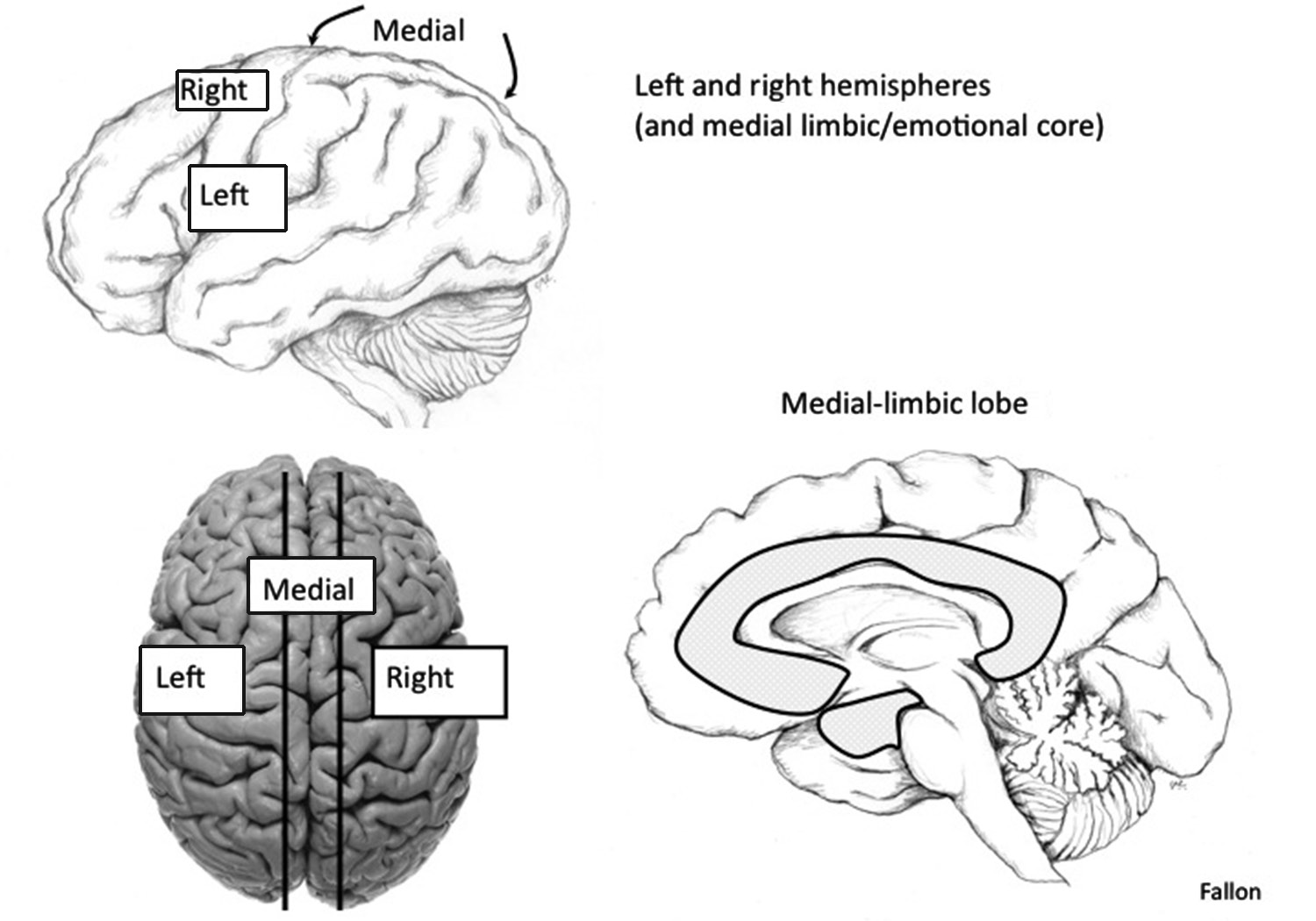
FIGURE 3B
: Brain hemispheres.
The next slicing of the Rubik's Cube brain is from front, or anterior, to back, or posterior. The most posterior region of the cortex is dedicated to the visual sensory system, as well as “association” cortices that have functions more complicated than simple seeing or touching or hearing, but rather cognitive tasks such as spatial processing. The external worldâup, down, left, right, close up, far awayâis mapped onto the cortex in the upper part of the posterior area, called the superior parietal cortex. People with damage to this brain area on one side will ignore the other half of their sensory world. So they may only perceive the numbers on the left side of a clock dial, but not the right side. Given a
blank circle, they will fill in the numbers on the dial from 1 to 12, but these will all be drawn on just one half of the clock. If the damage is done to the hemisphere that controls their nondominant hand, let's say the right superior parietal cortex for a right-hander (each hemisphere controls the opposite side of the body), then they will go the extra step in their “agnosia.” They will be able to move the opposite leg, and feel a pinch on that leg, but they may ask the doctor or nurse to remove the leg from the hospital bed because it is foreign and doesn't belong to their body.