The Spark of Life: Electricity in the Human Body (14 page)
Read The Spark of Life: Electricity in the Human Body Online
Authors: Frances Ashcroft

Muscle stiffness is worse when patients try to initiate a sudden movement after a period of rest, but gets gradually better with continued exercise. Even at rest, their muscles are continuously undergoing minute subthreshold contractions. Like horses with HYPP, it is as if they are doing continuous isometric exercises. Consequently, they tend to have a fine physique and very well-developed muscles – so much so that they can resemble body builders. Yet despite an athletic-looking figure, their muscles often let them down. One patient recalls that having crouched down in the typical starting position for a race, his muscles would immediately freeze when he stood up. He said, ‘My legs would lock into complete extension. It was like trying to run on stilts. I’d actually start running in maybe the last 20 or 30 metres.’
Goats Show the Way
The myotonic goats turned out to be the key to understanding myotonia congenita in humans. The Cincinnati physiologist Shirley Bryant had always been interested in muscle disorders. A brilliant and vivacious character who wore his hair in a ponytail even in his seventies, Bryant was a strong advocate of the idea that the study of animal diseases could elucidate similar human conditions, and he kept a strange menagerie of creatures, all of whom had inherited movement disorders, including tumbler pigeons, roller pigeons and a rare Australian marsupial, the Rottnest quokka. So when in the late 1950s he read of a herd of goats in Tennessee that fell down every time a train whistled past their field, he decided to investigate.

Bryant set off in a rented truck with an ex-convict driver. Initially, he had some difficulties in obtaining goats for his experiments. Fearing he would quickly complain that any fainting goats they sold him were defective and send them back, the farmers only gave him normal ones. Bryant had to make a second trip to Tennessee and it took some persuasion to reassure them it was the stiff-legged goats he actually wanted.
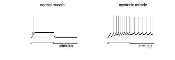
Once back in his laboratory, Bryant removed a small piece of muscle from between the ribs of the goats under anaesthesia. The operation is simple, painless and does not hurt the animal: indeed, many patients have undergone similar biopsies. Importantly, these intercostal muscles are very short and provide a tendon-to-tendon preparation that ensures the muscle is undamaged. Bryant first inserted a fine glass electrode into the cell to record the potential across the muscle fibre membrane, and then a second electrode to stimulate electrical activity. He observed that in a normal muscle fibre the application of a small positive current produced a single action potential and a single muscle twitch. However, in muscle fibres isolated from myotonic goats the same current produced a burst of impulses that sometimes continued long after the stimulus had stopped. In effect, they were firing without being signalled to do so. This caused the muscle to enter an extended period of contraction and explained why the goats’ legs became stiff and they fell over – their muscles were simply being excited more strongly. Similar findings were obtained using muscle biopsies from patients with myotonic congenita, showing that the human and goat diseases had a similar origin.
Unlike nerve fibres, muscle fibres have a high density of chloride channels, and in normal muscle the flow of chloride ions across the membrane dampens down electrical excitability, ensuring that a single nerve impulse produces only a single muscle twitch. Bryant conjectured that myotonic muscle might lack functional chloride channels, leading to enhanced excitability and sustained contraction. His experiments strongly supported this idea, although he was unable to measure the chloride current directly as there was no suitable voltage clamp for muscle at that time. Some years later, Richard Adrian, a muscle specialist at Cambridge University in England, invented a new tool that was exactly what Bryant needed to confirm his theory.

Myotonic muscle produces more electrical impulses when stimulated and these can continue even after the stimulus has stopped. If the electrical signal is played back through an audio amplifier, the normal muscle gives a single ‘click’ when stimulated, but the myotonic muscle sounds like an attacking dive-bomber.
In 1973, Bryant obtained permission to travel to England, together with four of his precious goats. It was not easy to take the goats to England because of fears that this might introduce a disease called bluetongue and a special Parliamentary Order, known as the ‘Importation of Goats Order No.1’, had to be passed to allow their importation. There was much media interest in the story, which was splashed across the front page of the
Wall Street Journal
. Despite this, the goats nearly did not make it. They were impounded at London’s Heathrow airport because they did not have their papers. Bryant had left for Cambridge in advance in order to be there to meet the goats, leaving his colleague to complete their shipping papers – but his colleague forgot to do so! With the goats in imminent danger of being shot, Bryant telephoned his Cambridge colleague. Roused from breakfast, Adrian used his very considerable persuasive powers to convince the authorities that the requirement to immediately destroy animals lacking the necessary papers on arrival strictly applied only to those that were ‘disembarking’ – and not those that were ‘disemplaning’. The goats were given a day’s grace, by which time, fortunately, their papers had arrived.
A perfectionist, and something of a procrastinator, Bryant never got around to publishing much of the data he and Adrian collected in Cambridge using the voltage clamp. It joined a mass of other unpublished data that filled a whole filing cabinet (visitors to his lab were often amazed when he showed them elegant experiments, conducted years earlier, that he had never got around to publishing, but which shed light on some current problem). Nevertheless, their studies clearly showed that myotonic muscle has a lower chloride current and that this is sufficient to explain the repetitive action potential firing characteristic of myotonia.
In 1992, the gene that codes for the human muscle chloride channel was sequenced, enabling people with myotonia congenita to be tested for mutations in the gene. Almost immediately, the first mutations were identified – and shortly after they were found in the descendants of Thomsen. To date, tens of other mutations have been described in the chloride channel gene, including the one responsible for myotonia in goats, and all of them result in a loss of function. Moreover, it turns out that muscle stiffness, like that characteristic of myotonia congenita, can also be produced by mutations in other ion channel genes. Thomsen’s myotonia, however, is of special scientific significance, as it was the first disorder to be linked to a defective ion channel. Many other ion channel diseases have been found subsequently, so many, in fact, that they have the distinction of having their own collective noun. They are known as the channelopathies.
Excitation–Contraction Coupling
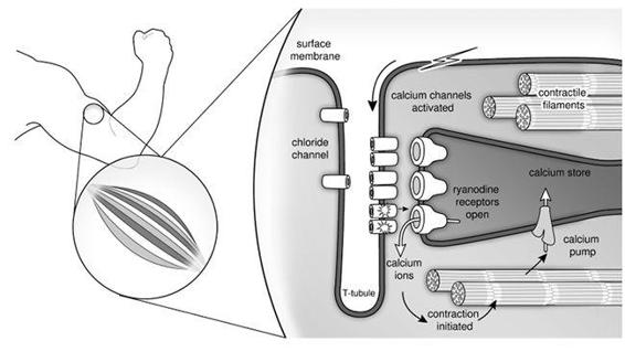
The question of how the muscle action potential stimulates a skeletal muscle fibre to contract has occupied scientists for centuries. We now know that muscle contraction is triggered by an increase in the intracellular concentration of calcium ions. At rest, the calcium concentration inside the muscle cell is very low. Electrical stimulation of the muscle causes a dramatic rise in calcium, which binds to the contractile proteins and leads to shortening of the muscle. However, the calcium ions do not come from outside the cell, but rather from a membrane-bound intracellular store called the sarcoplasmic reticulum. Calcium channels known as ryanodine receptors sit in the membrane of the sarcoplasmic reticulum and regulate the release of calcium. When they open, calcium floods out into the interior of the muscle fibre and triggers muscle contraction. When they close, calcium is quickly pumped back into the store, and the muscle relaxes. Ryanodine receptors are so called because they bind the plant alkaloid ryanodine with very high affinity.

The membranes of our muscles are full of ion channels. Calcium channels in the tubular membranes sense the voltage difference across the surface and tubular membranes and pass this information onto the ryanodine receptors, which sit in the intracellular membranes of the sarcoplasmic reticulum, the muscle’s calcium store. When the ryanodine receptors open, calcium rushes out, binds to the contractile filaments and causes the muscle to shorten. Muscle relaxation occurs when calcium is pumped back into the store, and its intracellular concentration falls. Chloride channels also sit in the surface and tubular membranes.
Precisely how the skeletal muscle action potential triggers the opening of the ryanodine receptors is still something of a mystery. After all, the action potential occurs in the surface membrane of the muscle and the ryanodine receptors are located in the membrane of the intracellular stores. Although these membranes are found in close apposition at specialized junctions, which lie within the tubular invaginations of the surface membrance, they never actually touch. It is clear, however, that voltage-sensitive calcium channels in the tubular membranes are somehow involved. One favoured idea is that the two kinds of calcium channel are in direct physical contact and that the ryanodine receptor channels, in effect, commandeer the tubular calcium channels’ voltage sensor. As a consequence, a muscle action potential opens the ryanodine receptors, causing calcium ions to rush out of the intracellular stores and trigger muscle contraction.
Shiver My Timbers
Mutations in the ryanodine receptors – the calcium release channels of the intracellular calcium stores – can also cause trouble. Malignant hyperthermia is a rare disorder that affects only a small number of people (around 1 in 20,000 adults), but it is the anaesthetist’s nightmare. It occurs when a susceptible patient is given a common anaesthetic gas, such as halothane, or certain types of muscle relaxant. These trigger spontaneous contractions of their skeletal muscles and a marked increase in muscle metabolism which increases heat production by the muscles and leads to a very rapid rise in body temperature – sometimes as much as 1°C every five minutes. In essence, the patients simply shiver themselves hot. An attack is a medical emergency as unless it is immediately treated, the increase in body temperature can be fatal. It is one of the leading causes of death from anaesthesia.
Malignant hyperthermia is also found in pigs, where it is known as porcine stress syndrome. It was once widespread in the UK pig population and was of considerable economic importance because not only do afflicted pigs often die, but the meat also becomes very pale, soft and unsaleable. As the name implies, the condition is triggered by various forms of stress including exercise, sex (in boars), parturition, being transported to market or simply by being kept in overcrowded conditions. It results from a mutation in the ryanodine receptor that causes the channels to become leaky so that the calcium concentration in the muscle cell rises, thereby stimulating metabolism, muscle contraction and an increase in body temperature. The skin of the animal becomes red and blotchy, and it may die of heat stoke within twenty minutes of the onset of an attack.
