The Spark of Life: Electricity in the Human Body (18 page)
Read The Spark of Life: Electricity in the Human Body Online
Authors: Frances Ashcroft

Speaking in Sparks
The discharges produced by electric fish can be grouped into two kinds: pulse-type and wave-type. Pulse-type electric fish like the elephant nose fish
Gnathonemus
emit a stream of brief electric pulses a few millivolts in amplitude. Wave-type electric fish such as the knifefish
Gymnarchus
emit a continuous electric current that oscillates in strength. These sinusoidal oscillations are extremely constant – almost as good as a commercial oscillator – and are produced at a frequency of 800 to 1,000 cycles per second.
Both types of fish are able to tune the frequency of their signals, which can vary not only between species and sexes, but also between individual fish. This provides a unique form of communication. The distinctive electrical pattern produced by different species of elephant fish, for example, enables them to detect others of the same species, an important consideration when finding a mate in dark and gloomy waters. Within a species, the frequency at which a fish emits is determined by its place in the social pecking order. The higher up in the hierarchy (i.e. the greater the status of the fish) the higher the frequency it uses. This is probably because it costs more energy to produce a higher frequency discharge so that only the ‘fittest’ fish can maintain their position in the hierarchy. It is the electrical equivalent of the peacock’s showy tail.
It is crucially important for a fish to be able to discriminate its own electric signal from those of others in the vicinity. Wave-type electric fish do this by generating signals at a fixed frequency. Each individual emits at its own frequency in much the same way as different radio stations broadcast at different frequencies. However, the number of frequencies is limited so that it sometimes happens that two fish which transmit at the same frequency meet up. This can cause a problem because it becomes unclear which signal arises from which fish, in the same way as it is difficult to distinguish two radio programmes broadcast at the same frequency. Essentially, the fish jam each other’s signal, thereby disrupting their electrolocation ability. Should this happen, the fish shift their frequencies relative to one another to maintain their privacy and avoid jamming each other’s signals. This separates out the signals emitted by individuals within communication range.
But it is not always sweetness and light and gentlemen’s agreements. In a fight, jamming your rival’s signal can disorientate it and give you a competitive advantage. Both male and female brown ghost knifefish appear to use such shock tactics when they come into conflict with a rival. Usually if they encounter another fish they switch their frequency to avoid interference, but if they are in competition they deliberately try to jam their opponent’s signal and establish dominance. In the social hierarchy of the brown ghost knife fish, the larger and more dominant males emit at a higher frequency and aggressively ramp up their electrical discharge frequency when they encounter a potential rival. This can result in a frequency war, with each fish trying to out-pitch the other’s electric signal and so disorientate its competitor.
Amorous male elephant nose fish also use electric signals to lure females. Different species of fish produce pulses that differ in magnitude, duration and frequency, and females are attuned to the signals produced by males of their own species. In some species, complex electrical courtship duets occur, analogous to the courtship songs of birds. Males of some nocturnal gymnotiform fish, for example, serenade their potential mates with long electrical hums and spawning elicits a frenzied electrical extravaganza. It is a costly concert for as much as 20 per cent of the energy consumed by the male fish is used to generate their electrical displays. These mega-signals serve to advertise the healthiest males, enabling a female to select the best mate. But this strategy brings a concomitant disadvantage. The electric signals are also picked up by electrosensitive predators, so that the males’ numbers are rapidly depleted and few male fish remain by the end of the mating season. To help prevent this decimation, male fish emit high frequency signals throughout the night, when females are more receptive and ready to spawn, but switch to low frequency songs during the day. Sexual strategies appear to be as intricately balanced in male electric fish as in their human counterparts.
7
The Heart of the Matter
Be still, my heart; thou hast known worse than this.
Homer
Early one summer morning, Alex was getting ready for school. Although she was anxious about her exams later that day, she was not unduly stressed and there was nothing that marked the day out as unusual. At least not until she went into the bathroom, reached out to turn on the light – and slid silently unconscious to the floor. Luckily, her mother saw it happen and rushed to the rescue. But this was no simple fainting fit. Alex had a serious heart problem and her increasingly frantic mother was unable to resuscitate her.
By chance, Alex lived close to a fire station and a local fireman picked up the emergency call. He quickly rushed to the rescue and administered cardiopulmonary resuscitation until the ambulance arrived, thus ensuring that her brain and other tissues were supplied with oxygen even though her heart was not working properly and she was no longer breathing. During the journey to the hospital her heart stopped and was restarted more than once. She was unconscious for seventeen hours but eventually recovered.
Subsequent analysis revealed that Alex has an abnormality in the electrical activity of her heart that predisposes her to blackouts and sudden cardiac death. It runs in the family. Her grandmother died in her sleep in her twenties, and her father suffered numerous fainting spells as a child and died young, just a year before Alex’s attack. It seems very likely that they carried the same genetic defect that Alex does.
Alex and her relatives are not alone. Other families have experienced similar tragedies, with one or more children or young adults dying in their sleep, when taking exercise, or when stressed. There are even tales of children suddenly collapsing when reprimanded by their teachers, or while running around in the playground. It is not an exaggeration to say that some children with this condition really have died of fright. Happily, our increased understanding of the electrical activity of the heart means that this disease can now be diagnosed from an electrocardiogram or by a simple genetic test, and it can also be successfully treated.
The Beat Goes On
It has been known for centuries that the heart has an intrinsic rhythm and can continue to beat when it is removed from the living animal. One of the first to describe the phenomenon was the great Roman physician Galen, and subsequently many others, including Leonardo da Vinci, reported that the heart moves by itself. William Harvey even showed that when the heart of an eel was cut into ever-smaller parts each individual piece continued to pulsate. This intrinsic activity may have inspired the classical Greek idea that the heart was the seat of the soul. However, the heartbeat has no spiritual origin, but derives instead from electrical events taking place within the cardiac cells themselves.
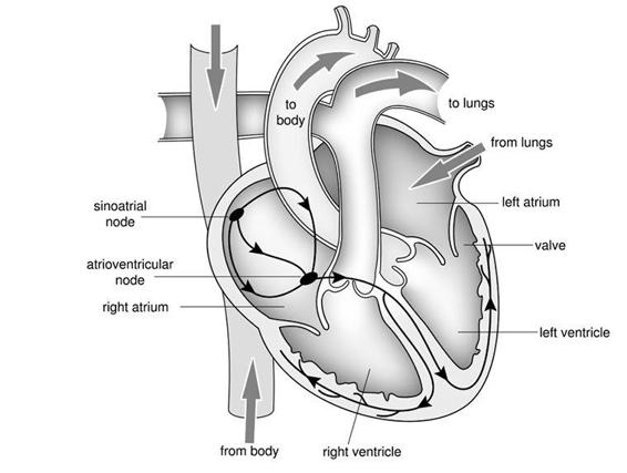
In essence, your heart is a pump that is controlled by electricity. Blood enters via the upper chambers (the atria), which contract first and force blood into the much larger lower chambers (the ventricles). The ventricles contract in synchrony about half a second later, the right ventricle pumping blood to the lungs and the left ventricle sending it around the body.
Non-return valves lie between the upper and lower chambers of the heart so that the blood only flows in one direction; from the atria to the ventricles. Similarly, non-return valves guard the exits from the ventricles into the great vessels. If these valves leak, as can happen with age, then blood is pumped less efficiently, so that the body receives less oxygen and you feel constantly tired. The chambers on the right and left side of the heart are physically separate which ensures that oxygen-rich blood coming from the lungs is not mixed with oxygen-depleted blood returning from the tissues. However, because heart cells are wired together, they contract in synchrony, so that the heart beats as a single organ.

The electrical system of the heart. The pacemaker cells lie within the sinus node in the wall of the right atrium. The black arrowed lines indicate the bundles of fibres that provide the path along which the electrical signals are conducted to the lower chambers (the ventricles). The two sides of the heart are physically separate but contract together. The pulmonary artery carries blood from the right side of the heart to the lungs. Having picked up oxygen there, blood is returned to the left side of the heart, which then pumps it into the aorta and around the body. The period when the heart is contracted is known as ‘systole’ and the time it is fully relaxed as ‘diastole’.
Each heartbeat originates in a group of pacemaker cells (known as the sinus node), which lie in the upper right chamber of the heart. These cells generate electrical impulses that are conducted to the rest of the heart along specialized pathways: first to the atrio-ventricular node, which lies at the junction between the right atrium and the ventricles, and then to the walls of the ventricles themselves. The time lag associated with electrical transmission ensures that the electrical signals reach the upper chambers before the lower ones, so that first the atria are triggered to contract, and then the ventricles. The timing of this spread of excitation is crucial for the ability of the heart to serve as a pump. If it is disrupted, the heart no longer beats regularly and its capacity to pump blood is compromised.
Although the average heart rate at rest is 70 beats a minute (that’s about 100,000 beats a day), it varies widely between individuals. Athletes have much lower resting heart rates, often as little as 40 beats a minute. One of the lowest ever recorded, a mere 28 beats per minute, was that of the cyclist Miguel Indurain, who won the Tour de France five times in a row. By contrast, babies’ hearts beat much faster than adults, speeding along at 130 to 150 beats a minute. It turns out that heart rate varies with body size, so that smaller animals (including babies) have higher resting heart rates: the heart of the tiny shrew races away at 600 beats a minute while that of the elephant can only manage a ponderous 25 beats a minute.
The Electrocardiogram
The electrical signals produced by heart cells give rise to tiny fluctuations in the electrical potential at the surface of the body that can be picked up by surface electrodes attached to the skin. This is the basis of the electrocardiogram, commonly abbreviated as ECG (or EKG in the United States).

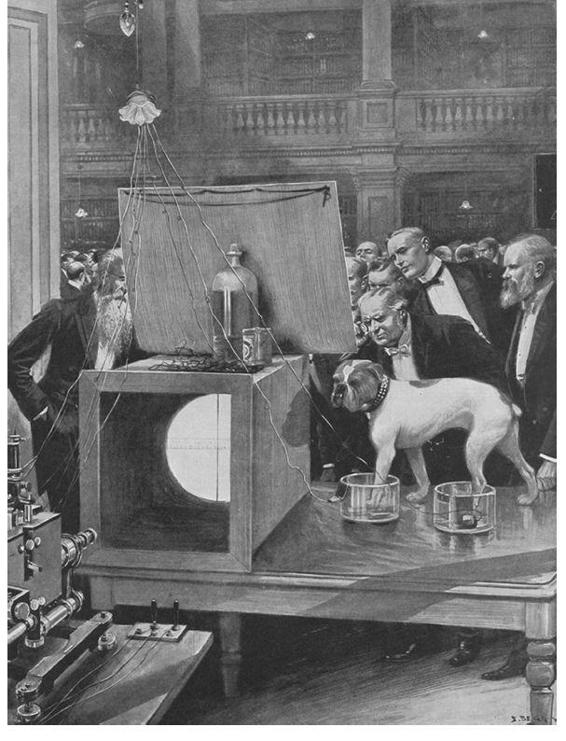
August Waller’s pet dog Jimmie was the most popular personage at the annual conversazione of the Royal Society at Burlington House. This scientific party for both scientists and the general public is still held and it traditionally includes many demonstrations. Jimmie stands sedately with his left paws in a conductive salt solution, which is connected to an Einthoven string galvanometer (the large box on the left) that measures his every heartbeat. The string is illuminated by limelight so that its shadow is projected on a sheet, and it vibrates with the bulldog’s heartbeat. The experiment is not painful, as many of the audience discovered when they volunteered to take Jimmie’s place. August Waller is seen on the far left.
The electrical activity of the heart was first recorded by Augustus Waller in 1887 both in himself and in his pet dog Jimmie. His demonstration of the method at the annual conversazione of the Royal Society of London in 1909, which was open to the public, was reported in the
Illustrated London News.
It triggered a storm of protest in Parliament, with Mr Ellis Griffith, the MP for Anglesey, demanding to know if the Cruelty to Animals Act of 1876 had been contravened.
The
Times
stated the Secretary of State, one Mr Gladstone,
1
replied, ‘I understand that the dog stood for some time in water, to which sodium chloride had been added, or, in other words, a little common salt. If my hon[ourable] friend has ever paddled in the sea, he will have understood the sensation. (Laughter.) The dog – a finely developed bulldog – was neither tied nor muzzled. He wore a leather collar ornamented with brass studs [this had been referred to by Mr. Griffith in far more emotive terms as “a leather strap with sharp nails [. . .] secured around the dog’s neck”]. Had the experiment been painful, the pain no doubt would have been more immediately felt by those nearest the dog. (Laughter.) There was no sign of this.’ He might have added that after Jimmie had shown the way, the ladies in the audience queued up to have their heartbeats recorded, by dipping their hands in pots of salt solution and ‘their hearts were in every case much steadier than Jimmie’s’. As this story also shows, the English concern about animal experimentation has a long history.
Waller’s early recordings were of poor quality and unsuitable for clinical purposes, and he is reputed to have said that he did not imagine electrocardiography was likely to find any very extensive use in the hospital and that, at most, it might be ‘of rare and occasional use to afford a concrete graphic record of some rare anomaly of cardiac action’. But technical innovations meant that by the 1920s it was routinely used to diagnose heart problems, and it remains an important clinical tool today.
The key was the development of very sensitive instruments, capable of detecting the tiny electric currents produced on the surface of the body when the heart beats. The pioneer in this field was Willem Einthoven, who won the Nobel Prize in 1924 for his invention of the string galvanometer. It consisted of a fine glass fibre, coated with silver to ensure that it could conduct current, which was suspended between two very strong electromagnets. When a current passed though the filament (the ‘string’ of the galvanometer), the electromagnetic field caused it to move. The greater the current, the more the filament was displaced. This tiny movement was detected by shining a light on the fibre, and the shadow it cast was recorded on a moving photographic plate. All that was needed was to connect the conductive filament to the body. This was done by attaching wires to each end of the filament and immersing the other end of the wires in pots of salt solution. Dipping the hands and feet in the solution completed the electrical connection between the ‘string’ and the skin. Current from the heart, picked up via the surface of the body, was then able to influence the movement of the filament.
The original string galvanometer was huge. It weighed several tons, took five people to operate and required constant running water to cool the electromagnets. The glass filament, however, had to be very light and thin. It was made by melting quartz glass in a crucible. This was then drawn out into a fine filament by a most unusual means – one more reminiscent of a Boy Scout improvisation than the conventional image of a scientific experiment. The molten glass was attached to an arrow that was then shot across the room, dragging the filament with it and stretching the glass into a very fine ‘string’. This was then coated with silver to make it electrically conductive. Safety considerations would undoubtedly ban this experiment today, but fortunately we now have other methods to record tiny currents.
Early photographs show Einthoven sitting with both hands and his left foot (trouser leg carefully rolled up) in separate tubs of conductive salt solution, which were wired up to the monitoring equipment. Today, conducting jelly is used to attach the recording electrodes, one on each arm and one on the left leg. The equipment has also got a lot smaller. Einthoven’s original machine occupied two rooms, but nowadays portable twenty-four-hour heart monitors are available that can be worn as the patient goes about their normal life.
The ECG simply reflects the sum of the electrical signals from individual heart cells and it provides a very good, non-invasive indicator of their function. Each ECG complex consists of an initial bump known as the ‘P wave’, followed by a much larger and sharper bipolar peak known as the ‘QRS complex’ and then, two to three hundred milliseconds later, by the smaller and slower ‘T wave’. The P wave corresponds to the electrical activity of the atrial cells, while the QRS and T waves reflect the beginning and end of the electrical impulse (the action potential) in the ventricular cells. Since these electrical signals drive muscle contraction, the P wave also signifies the contraction of the atria and the interval between the QRS and T waves indicates the duration of ventricular contraction. The delay between the P and Q waves is due to the length of time it takes for the electrical signal to spread from the atria to the ventricles, whereas the interval between the Q and T waves reflects the duration of the ventricular action potential. Why Einthoven should have chosen to name the ECG peaks after the middle letters of the alphabet remains something of a mystery.
