Breast Imaging: A Core Review (2 page)
Read Breast Imaging: A Core Review Online
Authors: Biren A. Shah,Sabala Mandava
Tags: #Medical, #Radiology; Radiotherapy & Nuclear Medicine, #Radiology & Nuclear Medicine

As my area of practice is predominantly breast imaging, I thought of putting together a bank of questions in this subspecialty that would cover the curriculum tested on the ABR Core Exam. I discussed the concept with my colleague, Sabala Mandava, who was also of a similar mind, and we decided to do a question book that would be geared toward residents preparing for the Core Exam, but can also be useful to any radiologist practicing Breast Imaging.
We were then fortunate to be able to enlist multiple colleagues who were interested in contributing to the book. As this book developed, I started thinking about similar books for the other subjects tested on the Core Exam. After several weeks of discussion with Jonathan Pine and Amy Dinkel, from Lippincott William & Wilkins, the concept of a series of books was born.
I am very pleased that the
Breast Imaging: A Core Review
is the first in The Core Review Series. There are multiple books such as Musculoskeletal Radiology, Neuroradiology, and others that are either currently being worked on or in the near future will be added to series. The philosophy for each book in the series is to review the important concepts tested with approximately 300 questions, in a format similar to the new ABR Core Exam.
As Series Editor of
The Core Review Series
, it has been a great source of pleasure to not only be an author of one of the books, but also to work with many outstanding colleagues across the country who contributed to the series. This series represents countless hours of work and involvement by many and it would not have come together without their participation.
My hope for this series is that it will prove to be a useful and comprehensive guide for all residents as well as fellows and practicing radiologists.
Biren A. Shah
Series Editor
| | PREFACE |
With the changing of the Boards format, these are uncertain times for radiology residents. The days of preparing for the oral boards with multiple reviews of image interpretation will likely change. Instead, the Boards are now geared to a more comprehensive understanding of disease processes, the physics behind image acquisition, quality control, and safety. There is a paucity of study resources available for residents.
With this in mind, we wanted to provide a guide for residents to be able to assess their knowledge and review the material in a format that would be similar to the Boards. The questions are divided into different sections, as per the ABR Core Exam Study Guide, so as to make it easy for the readers to work on particular topics as needed. There are mostly multiple-choice questions with some extended matching questions. Each question has a corresponding answer with an explanation of not only why a particular option is correct but also why the other options are incorrect. There are also references provided for each question for those who want to delve more deeply into a specific subject. This format is also useful for radiologists preparing for Maintenance of Certification (MOC).
There are multiple colleagues, some of whom are our past fellows, who contributed to this publication. This book could not have been finished without the efforts of all these people who took time from their busy lives to research, write, and submit material in a timely manner. Our heartfelt thanks to all of them.
Many thanks to the staff at LWW, Jonathan Pine, Amy Dinkel, Jeff Gunning, Sree Vidya Dhanvanthri, and Priscilla Crater for giving us this opportunity and guiding us along the way.
Last, but certainly not the least, we are grateful to our families, who have endured our long hours of work and kept us smiling throughout the process.
We hope that this book will serve as a useful tool for residents on their road to becoming Board-certified radiologists and will continue to be a reference in their future careers.
Biren A. Shah, MD
Sabala R. Mandava, MD
| | CONTENTS |
Contributors
Series Foreword
Preface
1 Regulatory/Standards of Care
2 Breast Cancer Screening
3 Diagnostic Breast Imaging, Breast Pathology, and Breast Imaging Findings
4 Breast Intervention
5 Physics Related to Breast Imaging
Index
| 1 | Regulatory/Standards of Care |
QUESTIONS
1
Which of the following is a Mammography Quality Standards Act (MQSA) requirement for interpreting physicians?
A. 15 category 1 continuing medical education (CME) credits per year
B. 10 hours of initial new modality training (e.g., digital mammography)
C. Initial experience of 240 exams under direct supervision in the 6 months before starting to interpret mammography
D. Continuing experience of interpretation of 960 exams/12 months
2
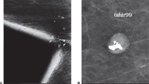
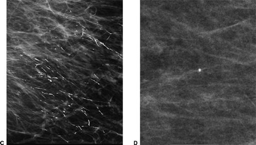
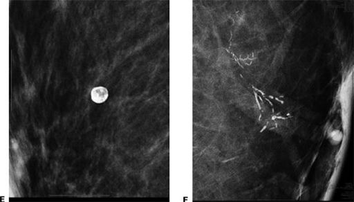
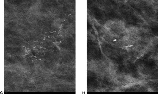
For each diagnostic image, below, assign the likely BI-RADS assessment of either BI-RADS 2 (answer choice “A”) or BI-RADS 4 (answer choice “B”). Each option may be used once, more than once, or not at all:


3
The approximate expected number of cancers that should be found in 1,000 initial screening mammograms is
A. 1 to 2
B. 6 to 10
C. 11 to 14
D. 15 to 19
E. 20 to 24
4
Over a year, 100 cancers are identified; 94 of these were identified based on biopsy recommendations from a screening mammogram and an additional 6 cancers developed after a negative mammogram. What is the sensitivity in this population?
A. 6%
B. 88%
C. 90%
D. 94%
E. 96%
5
When assessing for accurate positioning on mediolateral oblique (MLO) view, which of the following is correct?
A. A large amount of the upper abdomen should be visible.
B. The breast should be pulled out and down.
C. The pectoral muscle should widen at the axilla and extend to the nipple, and the anterior margin should be convex.
D. The inframammary fold should be neutral in position.
6
A patient has a negative screening mammogram study and 8 months later develops a palpable mass that is biopsied to reveal invasive ductal carcinoma. This is termed a
A. False negative
B. False positive
C. True positive
D. True negative
7
Which of the following quality control tests are performed weekly for filmscreen mammography?
A. Darkroom cleanliness
B. Processor quality control
C. Screen cleanliness
D. Viewbox cleanliness
E. Fixer retention
8a
An 85-year-old female with history of left mastectomy. The patient presented for a screening mammogram of the right breast. A radiopaque marker was placed on the nipple. Images are provided below.

