Phantoms in the Brain: Probing the Mysteries of the Human Mind (7 page)
Read Phantoms in the Brain: Probing the Mysteries of the Human Mind Online
Authors: V. S. Ramachandran,Sandra Blakeslee
Tags: #Medical, #Neurology, #Neuroscience

Of the many strange images that have remained with me from my medical school days, perhaps none is more vivid than that of the deformed little man you see in Figure 2.1 draped across the surface of the cerebral cortex—the so−called Penfield homunculus. The homunculus is the artist's whimsical depiction of the manner in which different points on the body surface are mapped onto the surface of the brain—the grotesquely deformed features are an attempt to indicate that certain body parts such as the lips and tongue are grossly overrepresented.
The map was drawn from information gleaned from real human brains. During the 1940s and 1950s, the brilliant Canadian neurosurgeon Wilder Penfield performed extensive brain surgeries on patients under local anesthetic (there are no pain receptors in the brain, even though it is a mass of nerve tissue). Often, much of the brain was exposed during the operation and Penfield seized this opportunity to do experiments that had never been tried before. He stimulated specific regions of the patients' brains with an electrode and simply asked them what they felt. All kinds of sensations, images, and even memories were elicited by the electrode and the areas of the brain that were responsible could be mapped.
Among other things, Penfield found a narrow strip running from top to bottom down both sides of the brain where his electrode produced sensations localized in various parts of the body. Up at the top of the brain, in the crevice that separates the two hemispheres, electrical stimulation elicited sensations in the genitals. Nearby stimuli evoked sensa−
Figure 2.1
(a) The representation of the body surface on the surface of the human brain (as discovered by
Wilder Penfield) behind the central sulcus. There are many such maps, but for clarity only one is shown here.
The homunculus ("little man") is upside down for the most part, and his feet are tucked onto the medial
surface (inner surface) of the parietal lobe near the very top, whereas the face is down near the bottom of the
outer surface. The face and hand occupy a disproportionately large share of the map. Notice, also that the
face area is below the hand area instead of being where it should
—
near the neck
—
and that the genitals are
represented below the foot. Could this provide an anatomical explanation of foot fetishes'? (b) A whimsical
24
three−dimensional model of the Penfield homunculus
—
the little man in the brain
—
depicting the
representation of body parts. Notice the gross overrepresentation of mouth and hands.
Reprinted with permission from the British Museum, London.
tions in the feet. As Penfield followed this strip down from the top of the brain, he discovered areas that receive sensations from the legs and trunk, from the hand (a large region with a very prominent representation of the thumb), the face, the lips and finally the thorax and voicebox. This "sensory homunculus," as it is now called, forms a greatly distorted representation of the body on the surface of the brain, with the parts that are particularly important taking up disproportionately large areas. For example, the area involved with the lips or with the fingers takes up as much space as the area involved with the entire trunk of the body. This is presumably because your lips and fingers are highly sensitive to touch and are capable of very fine discrimination, whereas your trunk is considerably less sensitive, requiring less cortical space. For the most part, the map is orderly though upside down: The foot is represented at the top and the outstretched arms are at the bottom. However, upon close
examination, you will see that the map is not entirely continuous. The face is not near the neck, where it should be, but is below the hand. The genitals, instead of being between the thighs, are located below the foot.4
These areas can be mapped out with even greater precision in other animals, particularly in monkeys. The researcher inserts a long thin needle made of steel or tungsten into the monkey's somatosensory cortex—the strip of brain tissue described earlier. If the needle tip comes to lie right next to the cell body of a neuron and if that neuron is active, it will generate tiny electrical currents that are picked up by the needle electrode and amplified. The signal can be displayed on an oscilloscope, making it possible to monitor the activity of that neuron.
For example, if you put an electrode into the monkey's somatosensory cortex and touch the monkey on a specific part of its body, the cell will fire. Each cell has its territory on the body surface—its own small patch of skin, so to speak—to which it responds. We call this the cell's receptive field. A map of the entire body surface exists in the brain, with each half of the body mapped onto the opposite side of the brain.
While animals are logical experimental subjects in which to examine the detailed structure and function of the brain's sensory regions, they have one obvious problem: Monkeys can't talk. Therefore, they cannot tell the experimenter, as Penfield's patients could, what they are feeling. Thus a large and important dimension is lost when animals are used in such experiments.
But despite this obvious limitation, a great deal can be learned by doing the right kinds of experiments. For instance, as we've noted, one important question concerns nature versus nurture: Are these body maps on the surface of the brain fixed, or can they change with experience as we grow from newborns to infancy, through adolescence and into old age? And even if the maps are already there at birth, to what extent can they be modified in the adult?5
It was these questions that prompted Tim Pons and his colleagues to embark on their research. Their strategy was to record signals from the brains of monkeys who had undergone dorsal rhizotomy—a procedure in which all the nerve fibers carrying sensory information from one arm into the spinal cord are completely severed.6 Eleven years after the surgery, they anesthetized the animals, opened their skulls and recorded from the somatosensory map. Since the monkey's paralyzed arm was not sending messages to the brain, you would not expect to record any sig−
nals when you touch the monkey's useless hand and record from the "hand area" of the brain. There should be a big patch of silent cortex corresponding to the affected hand.
25
Indeed, when the researchers stroked the useless hand, there was no activity in this region. But to their amazement they found that when they touched the monkey's face, the cells in the brain corresponding to the
"dead" hand started firing vigorously. (So did cells corresponding to the face, but those were expected to fire.) It appeared that sensory information from the monkey's face not only went to the face area of the cortex, as it would in a normal animal, but it had also invaded the territory of the paralyzed hand!
The implications of this finding are astonishing: It means that you
can
change the map; you can alter the brain circuitry of an adult animal, and connections can be modified over distances spanning a centimeter or more.
Upon reading Pons's paper, I thought, "My God! Might this be an explanation for phantom limbs?" What did the monkey actually "feel" when its face was being stroked? Since its "hand" cortex was also being excited, did it perceive sensations as arising from the useless hand as well as the face? Or would it use higher brain centers to reinterpret the sensations correctly as arising from the face alone? The monkey of course was silent on the subject.
It takes years to train a monkey to carry out even very simple tasks, let alone signal what part of its body is being touched. Then it occurred to me that you don't have to use a monkey. Why not answer the same question by touching the face of a human patient who has lost an arm? I telephoned my colleagues Dr. Mark Johnson and Dr. Rita Finkelstein in orthopedic surgery and asked, "Do you have any patients who have recently lost an arm?"
That is how I came to meet Tom. I called him up right away and asked whether he would like to participate in a study. Although initially shy and reticent in his mannerisms, Tom soon became eager to participate in our experiment. I was careful not to tell him what we hoped to find, so as not to bias his responses. Even though he was distressed by "itching" and painful sensations in his phantom fingers, he was cheerful, apparently pleased that he had survived the accident.
With Tom seated comfortably in my basement laboratory, I placed a blindfold over his eyes because I didn't want him to see where I was touching him. Then I took an ordinary Q−tip and started stroking various parts of his body surface, asking him to tell me where he felt the sensations. (My graduate student, who was watching, thought I was crazy.)
I swabbed his cheek. "What do you feel?"
"You are touching my cheek."
"Anything else?"
"Hey, you know it's funny," said Tom. "You're touching my missing thumb, my phantom thumb."
I moved the Q−tip to his upper lip. "How about here?"
"You're touching my index finger. And my upper lip."
"Really? Are you sure?"
"Yes. I can feel it both places."
"How about here?" I stroked his lower jaw with the swab.
26

cluster of points on the face that evoke sensations in the phantom and a second cluster on the upper arm, corresponding to the two body parts that are represented on either side (above and below) of the hand representation in the brain.8
It's not often in science (especially neurology) that you can make a simple prediction like this and confirm it with a few minutes of exploration using a Q−tip. The existence of two clusters of points suggests strongly that remapping of the kind seen in Pons's monkeys also occurs in the human brain. But there was still a nagging doubt: How can we
"Knowing Where to Scratch" / 31
be sure that such changes are actually taking place—that the map is really changing in people like Tom? To obtain more direct proof, we took advantage of a modern neuroimaging technique called magnetoence−phalography (MEG), which relies on the principle that if you touch different body parts, the localized electrical activity evoked in the Penfield map can be measured as changes in magnetic fields on the scalp. The major advantage of the technique is that it is noninvasive; one does not have to open the patient's scalp to peer inside the brain.
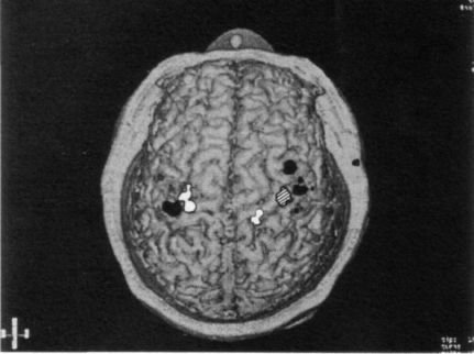
Using MEG, it is relatively easy in just a two−hour session to map out the entire body surface on the brain surface of any person willing to sit under the magnet. Not surprisingly, the map that results is quite similar to the original Penfield homunculus map, and there is very little variation from person to person in the gross layout of the map. When we conducted MEGs on four arm amputees, however, we found that the maps had changed over large distances, just as we had predicted. For example, a glance at Figure 2.3 reveals that the hand area (hatched) is missing in the right hemisphere and has been invaded by the sensory input from the face (in white) and upper arm (in gray). These observations, which I made in collaboration with a medical student, Tony Yang, and the neurologists Chris Gallen and Floyd Bloom, were in fact the first direct demonstration that such large−scale changes in the organization of the brain could occur in adult humans.
The implications are staggering. First and foremost, they suggest that brain maps can change, sometimes with astonishing rapidity. This finding flatly contradicts one of the most widely accepted dogmas in neurology—
the fixed nature of connections in the adult human brain. It had always been assumed that once this circuitry, including the Penfield map, has been laid down in fetal life or in early infancy, there is very little one can do to modify it in adulthood. Indeed, this presumed absence of plasticity in the adult brain is often invoked to explain why there is so little recovery of function after brain injury and why neurological ailments are so notoriously difficult to treat. But the evidence from Tom shows— contrary to what is taught in textbooks—that new, highly precise and functionally effective pathways can emerge in the adult brain as early as four weeks after injury. It certainly doesn't follow that revolutionary new treatments for neurological syndromes will emerge from this discovery right away, but it does provide some grounds for optimism.
Second, the findings may help explain the very existence of phantom limbs. The most popular medical explanation, noted earlier, is that nerves that once supplied the hand begin to innervate the stump. Moreover, 28