Ross & Wilson Anatomy and Physiology in Health and Illness (100 page)
Read Ross & Wilson Anatomy and Physiology in Health and Illness Online
Authors: Anne Waugh,Allison Grant
Tags: #Medical, #Nursing, #General, #Anatomy

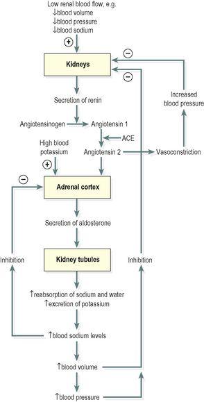
The blood potassium level regulates the amount of aldosterone produced by the adrenal cortex. When blood potassium levels rise, more aldosterone is secreted (
Fig. 9.11
). Low blood potassium has the opposite effect.
Angiotensin
(see below) also stimulates the release of aldosterone.
Figure 9.11
Negative feedback regulation of aldosterone secretion.
Renin–angiotensin–aldosterone system
When renal blood flow is reduced or blood sodium levels fall, the enzyme
renin
is secreted by kidney cells. Renin converts the plasma protein
angiotensinogen
, produced by the liver, to
angiotensin 1
. Angiotensin converting enzyme (ACE), formed in small quantities in the lungs, proximal kidney tubules and other tissues, converts angiotensin 1 to
angiotensin 2
, which stimulates secretion of aldosterone (
Fig. 9.11
). It also causes vasoconstriction and increases blood pressure.
Sex hormones
Sex hormones secreted by the adrenal cortex are mainly
androgens
(male sex hormones) and the amounts produced are insignificant compared with those secreted by the testes and ovaries in late puberty and adulthood (see
Ch. 18
).
Adrenal medulla
The medulla is completely surrounded by the adrenal cortex. It develops from nervous tissue in the embryo and is part of the sympathetic division of the autonomic nervous system (
Ch. 7
). It is stimulated by its extensive sympathetic nerve supply to produce the hormones
adrenaline
(epinephrine) and
noradrenaline
(norepinephrine).
Adrenaline (epinephrine) and noradrenaline (norepinephrine)
Noradrenaline is the postganglionic neurotransmitter of the sympathetic division of the autonomic nervous system (see
Fig. 7.46, p. 168
). Adrenaline and some noradrenaline are released into the blood from the adrenal medulla during stimulation of the sympathetic nervous system (see
Fig. 7.47, p. 169
). The action of these hormones prolongs and augments stimulation of the sympathetic nervous system. They are structurally very similar and this explains their similar effects. Together they potentiate the fight or flight response by:
•
increasing heart rate
•
increasing blood pressure
•
diverting blood to essential organs, including the heart, brain and skeletal muscles, by dilating their blood vessels and constricting those of less essential organs, such as the skin
•
increasing metabolic rate
•
dilating the pupils.
Adrenaline has a greater effect on the heart and metabolic processes whereas noradrenaline has more influence on blood vessels.
Response to stress
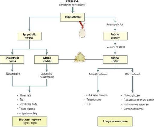
When the body is under stress homeostasis is disturbed. To restore it and, in some cases, to maintain life there are immediate and, if necessary, longer-term responses. Stressors include exercise, fasting, fright, temperature changes, infection, disease and emotional situations.
The
immediate response
is sometimes described as preparing for ‘fight or flight’. This is mediated by the sympathetic part of the autonomic nervous system and the principal effects are shown in
Figure 9.12
.
Figure 9.12
Responses to stressors that threaten homeostasis.
CRH = corticotrophin releasing hormone. ACTH = adrenocorticotrophic hormone.
In the
longer term
, ACTH from the anterior pituitary stimulates the release of glucocorticoids and mineralocorticoids from the adrenal cortex and a more prolonged response to stress occurs (
Fig. 9.12
).
Pancreatic islets
Learning outcomes
After studying this section you should be able to:
list the hormones secreted by the endocrine pancreas
describe the actions of insulin and glucagon
explain how blood glucose levels are regulated.
The structure of the pancreas is described in
Chapter 12
. The cells that make up the pancreatic islets (islets of Langerhans) are found in clusters irregularly distributed throughout the substance of the pancreas. Unlike the exocrine pancreas, which produces pancreatic juice (
p. 300
), there are no ducts leading from the clusters of islet cells. Pancreatic hormones are secreted directly into the bloodstream and circulate throughout the body.
There are three main types of cells in the pancreatic islets:
•
α (alpha) cells, which secrete
glucagon
•
β (beta) cells, which are the most numerous, secrete
insulin
•
δ (delta) cells, which secrete
somatostatin
(GHRIH,
pp. 210
and
219
).
The normal blood glucose level is between 3.5 and 8 mmol/litre (63 to 144 mg/100 ml). Blood glucose levels are controlled mainly by the opposing actions of insulin and glucagon:
•
glucagon increases blood glucose levels
•
insulin reduces blood glucose levels.
Insulin
Insulin is a polypeptide consisting of about 50 amino acids. Its main function is to lower raised blood nutrient levels, not only glucose but also amino acids and fatty acids. These effects are described as anabolic, i.e. they promote storage of nutrients. When these nutrients, especially glucose, are in excess of immediate needs insulin promotes their storage by:
•
acting on cell membranes and stimulating uptake and use of glucose by muscle and connective tissue cells
•
increasing conversion of glucose to glycogen (glycogenesis), especially in the liver and skeletal muscles
•
accelerating uptake of amino acids by cells, and the synthesis of protein


