Ross & Wilson Anatomy and Physiology in Health and Illness (96 page)
Read Ross & Wilson Anatomy and Physiology in Health and Illness Online
Authors: Anne Waugh,Allison Grant
Tags: #Medical, #Nursing, #General, #Anatomy

Figure 9.2
The position of the pituitary gland and its associated structures.
Blood supply
Arterial blood
This is supplied by branches from the internal carotid artery. The anterior lobe is supplied indirectly by blood that has already passed through a capillary bed in the hypothalamus but the posterior lobe is supplied directly.
Venous drainage
This comes from both lobes, containing hormones, and leaves the gland in short veins that enter the venous sinuses between the layers of dura mater.
The influence of the hypothalamus on the pituitary gland
The influence of the hypothalamus on the release of hormones is different in the anterior and posterior lobes of the pituitary gland.
The anterior pituitary
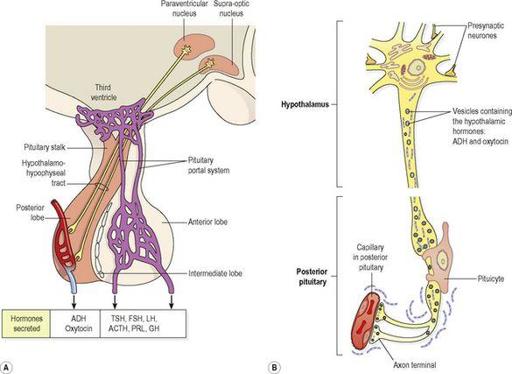
This is supplied indirectly with arterial blood that has already passed through a capillary bed in the hypothalamus (
Fig. 9.3A
). This network of blood vessels forms part of the
pituitary portal system
, which transports blood from the hypothalamus to the anterior pituitary where it enters thin-walled sinusoids that are in close contact with the secretory cells. As well as providing oxygen and nutrients, this blood transports
releasing
and
inhibiting hormones
secreted by the hypothalamus. These hormones influence secretion and release of other hormones formed in the anterior pituitary. The releasing and inhibiting hormones that stimulate and inhibit secretion of specific anterior pituitary hormones are shown in
Table 9.1
.
Figure 9.3
The pituitary gland. A.
The lobes of the pituitary gland and their relationship with the hypothalamus.
B.
Synthesis and storage of antidiuretic hormone and oxytocin.
Table 9.1
Hormones of the hypothalamus, anterior pituitary and their target tissues
| Hypothalamus | Anterior pituitary | Target gland or tissue |
|---|---|---|
| GHRH | GH | Most tissues |
| | | Many organs |
| GHRIH | GH inhibition | Thyroid gland |
| | TSH inhibition | Pancreatic islets |
| | | Most tissues |
| TRH | TSH | Thyroid gland |
| CRH | ACTH | Adrenal cortex |
| PRH | PRL | Breast |
| PIH | PRL inhibition | Breast |
| LHRH or | FSH | Ovaries and testes |
| GnRH | LH | Ovaries and testes |
GHRH = growth hormone releasing hormone
GH = growth hormone (somatotrophin)
GHRIH = growth hormone release inhibiting hormone (somatostatin)
TRH = thyrotrophin releasing hormone
TSH = thyroid stimulating hormone
CRH = corticotrophin releasing hormone
ACTH = adrenocorticotrophic hormone
PRH = prolactin releasing hormone
PRL = prolactin (lactogenic hormone)
PIH = prolactin inhibiting hormone (dopamine)
LHRH = luteinising hormone releasing hormone
GnRH = gonadotrophin releasing hormone
FSH = follicle stimulating hormone
LH = luteinising hormone
The posterior pituitary
This is formed from nervous tissue and consists of nerve cells surrounded by supporting cells called
pituicytes
. These neurones have their cell bodies in the supraoptic and paraventricular nuclei of the hypothalamus and their axons form a bundle known as the
hypothalamohypophyseal tract
(
Fig. 9.3A
). Posterior pituitary hormones are synthesised in the nerve cell bodies, transported along the axons and stored in vesicles within the axon terminals in the posterior pituitary (
Fig. 9.3B
). Their release by exocytosis is triggered by nerve impulses from the hypothalamus.
Anterior pituitary
Some of the hormones secreted by the anterior lobe stimulate or inhibit secretion by other endocrine glands (target glands) while others have a direct effect on target tissues.
Table 9.1
summarises the main relationships between the hormones of the hypothalamus, the anterior pituitary and target glands or tissues.
The release of an anterior pituitary hormone follows stimulation of the gland by a specific
releasing hormone
produced by the hypothalamus and carried to the gland through the pituitary portal system of blood vessels. The whole system is controlled by a
negative feedback mechanism
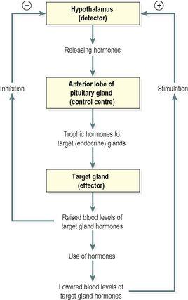
. That is, when there is a low level of a hormone in the blood supplying the hypothalamus it produces the appropriate releasing hormone that stimulates release of a
trophic hormone
by the anterior pituitary. This in turn stimulates the target gland to produce and release its hormone. As a result the blood level of that hormone rises and inhibits the secretion of releasing factor by the hypothalamus (
Fig. 9.4
).
Figure 9.4
Negative feedback regulation of secretion of hormones by the anterior lobe of the pituitary gland.
Growth hormone (GH)
This is the most abundant hormone synthesised by the anterior pituitary. It stimulates growth and division of most body cells but especially those in the bones and skeletal muscles. Body growth in response to the secretion of GH is evident during childhood and adolescence, and thereafter secretion of GH maintains the mass of bones and skeletal muscles. It also regulates aspects of metabolism in many organs, e.g. liver, intestines and pancreas; stimulates protein synthesis, especially tissue growth and repair; promotes breakdown of fats; and increases blood glucose levels (see
Ch. 12
).
Its release is stimulated by
growth hormone releasing hormone
(GHRH) and suppressed by
growth hormone release inhibiting hormone
(GHRIH), also known as
somatostatin
, both of which are secreted by the hypothalamus. Secretion of GH is greater at night during sleep and is also stimulated by hypoglycaemia, exercise and anxiety. The daily amount secreted peaks in adolescence and then declines with age. Inhibition of GH secretion occurs by a negative feedback mechanism when the blood level rises and also when GHRIH is released by the hypothalamus. GHRIH also suppresses secretion of TSH and gastrointestinal secretions, e.g. gastric juice, gastrin and cholecystokinin (see
Ch. 12
).
Thyroid stimulating hormone (TSH)
This hormone is synthesised by the anterior pituitary and its release is stimulated by thyrotrophin releasing hormone (TRH) from the hypothalamus. It stimulates growth and activity of the thyroid gland, which secretes the hormones
thyroxine
(T
4
) and
tri-iodothyronine
(T
3
). Release is lowest in the early evening and highest during the night. Secretion is regulated by a negative feedback mechanism (
Fig. 9.4
). When the blood level of thyroid hormones is high, secretion of TSH is reduced, and vice versa.
Adrenocorticotrophic hormone (ACTH, corticotrophin)
Corticotrophin releasing hormone (CRH) from the hypothalamus promotes the synthesis and release of ACTH by the anterior pituitary. This increases the concentration of cholesterol and steroids within the adrenal cortex and the output of steroid hormones, especially
cortisol
.
ACTH levels are highest at about 8 a.m. and fall to their lowest about midnight, although high levels sometimes occur at midday and 6 p.m. This circadian rhythm is maintained throughout life. It is associated with the sleep pattern and adjustment to changes takes several days, following, e.g., changing work shifts, travelling to a different time zone (jet lag).
Secretion is also regulated by a negative feedback mechanism, being suppressed when the blood level of ACTH rises (
Fig. 9.4
). Other factors that stimulate secretion include hypoglycaemia, exercise and other stressors, e.g. emotional states and fever.
Prolactin
This hormone is secreted during pregnancy to prepare the breasts for
lactation
(milk production) after childbirth. The blood level of prolactin is stimulated by prolactin releasing hormone (PRH) released from the hypothalamus and it is lowered by prolactin inhibiting hormone (PIH,
dopamine
) and by an increased blood level of prolactin. Immediately after birth, suckling stimulates prolactin secretion and lactation. The resultant high blood level is a factor in reducing the incidence of conception during lactation.
Prolactin, together with oestrogens, corticosteroids, insulin and thyroxine, is involved in initiating and maintaining lactation. Prolactin secretion is related to sleep, i.e. it is raised during any period of sleep, night or day.
Gonadotrophins
Just before puberty two gonadotrophins (sex hormones) are secreted in gradually increasing amounts by the anterior pituitary in response to
luteinising hormone releasing hormone
(LHRH), also known as
gonadotrophin releasing hormone
(GnRH). At puberty secretion increases, further enabling normal adult functioning of the reproductive organs; in both males and females the hormones responsible are:
•
follicle stimulating hormone (FSH)
•
luteinising hormone (LH).
In both sexes
FSH stimulates production of gametes (ova or spermatozoa) by the gonads.
In females
LH and FSH are involved in secretion of the hormones
oestrogen
and
progesterone
during the menstrual cycle (see
Figs 18.9
and
18.10, p. 444
). As the levels of oestrogen and progesterone rise, secretion of LH and FSH is suppressed.
In males
LH, also called interstitial cell stimulating hormone (ICSH) stimulates the interstitial cells of the testes to secrete the hormone
testosterone
(see
Ch. 18
).
Table 9.2
summarises the hormonal secretions of the anterior pituitary.
Table 9.2
Summary of the hormones secreted by the anterior pituitary gland and their functions
| Hormone | Function |
|---|---|
| Growth hormone (GH) | Regulates metabolism, promotes tissue growth especially of bones and muscles |
| Thyroid stimulating hormone (TSH) | Stimulates growth and activity of thyroid gland and secretion of T 3 and T 4 |
| Adrenocorticotrophic hormone (ACTH) | Stimulates the adrenal cortex to secrete glucocorticoids |
| Prolactin (PRL) | Stimulates milk production in the breasts |
| Follicle stimulating hormone (FSH) | Stimulates production of sperm in the testes, stimulates secretion of oestrogen by the ovaries, maturation of ovarian follicles, ovulation |
| Luteinising hormone (LH) | Stimulates secretion of testosterone by the testes, stimulates secretion of progesterone by the corpus luteum |


