Ross & Wilson Anatomy and Physiology in Health and Illness (181 page)
Read Ross & Wilson Anatomy and Physiology in Health and Illness Online
Authors: Anne Waugh,Allison Grant
Tags: #Medical, #Nursing, #General, #Anatomy

•
1 frontal bone
•
2 parietal bones
•
2 temporal bones
•
1 occipital bone
•
1 sphenoid bone
•
1 ethmoid bone.
Frontal bone
This is the bone of the forehead. It forms part of the
orbital cavities
(eye sockets) and the prominent ridges above the eyes, the
supraorbital margins
. Just above the supraorbital margins, within the bone, are two air-filled cavities or
sinuses
lined with ciliated mucous membrane, which open into the nasal cavity.
The
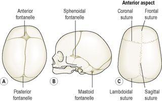
coronal suture
joins the frontal and parietal bones and other fibrous joints are formed with the sphenoid, zygomatic, lacrimal, nasal and ethmoid bones. The bone originates in two parts joined in the midline by the
frontal suture
(see
Fig. 16.18
).
Figure 16.18
The skull showing the fontanelles and sutures. A.
Fontanelles viewed from above.
B.
Fontanelles viewed from the side.
C.
Main sutures viewed from above when ossification is complete.
Parietal bones
These bones form the sides and roof of the skull. They articulate with each other at the
sagittal suture
, with the frontal bone at the coronal suture, with the occipital bone at the
lambdoidal suture
and with the temporal bones at the
squamous sutures
. The inner surface is concave and is grooved to accommodate the brain and blood vessels.
Temporal bones (
Fig. 16.12
)
These bones lie one on each side of the head and form fibrous immovable joints with the parietal, occipital, sphenoid and zygomatic bones. Each temporal bone has several important features.
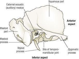
Figure 16.12
The right temporal bone.
Lateral view.
The
squamous part
is the thin fan-shaped area that articulates with the parietal bone. The
zygomatic process
articulates with the zygomatic bone to form the zygomatic arch (cheekbone).
The
mastoid part
contains the mastoid process, a thickened region easily felt behind the ear. It contains a large number of very small air sinuses that communicate with the middle ear and are lined with squamous epithelium.
The
petrous portion
forms part of the base of the skull and contains the organs of hearing (the spiral organ) and balance.
The temporal bone articulates with the mandible at the
temporomandibular joint
, the only movable joint of the skull. Immediately behind this articulating surface is the
external acoustic meatus
(auditory canal), which passes inwards towards the petrous portion of the bone.
The styloid process projects from the lower process of the temporal bone, and supports the hyoid bone and muscles associated with the tongue and pharynx.
Occipital bone (
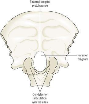
Fig. 16.13
)
This bone forms the back of the head and part of the base of the skull. It has immovable fibrous joints with the parietal, temporal and sphenoid bones. Its inner surface is deeply concave and the concavity is occupied by the occipital lobes of the cerebrum and by the cerebellum. The occiput has two articular condyles that form condyloid joints (
p. 404
) with the first bone of the vertebral column, the atlas. This joint permits nodding movements of the head. Between the condyles is the
foramen magnum
(meaning ‘large hole’) through which the spinal cord passes into the cranial cavity.
Figure 16.13
The occipital bone
– viewed from below.
Sphenoid bone (
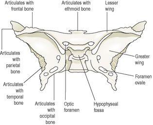
Fig. 16.14
)
This bone occupies the middle portion of the base of the skull and it articulates with the occipital, temporal, parietal and frontal bones (
Fig. 16.11
). It links the cranial and facial bones, and cross-braces the skull. On the superior surface in the middle of the bone is a little saddle-shaped depression, the
hypophyseal fossa
(
sella turcica
) in which the
pituitary gland
rests. The body of the bone contains some fairly large air sinuses lined with ciliated mucous membrane with openings into the nasal cavity.
Figure 16.14
The sphenoid bone
– viewed from above.
Ethmoid bone (
Fig. 16.15
)
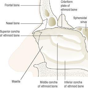
The ethmoid bone occupies the anterior part of the base of the skull and helps to form the orbital cavity, the nasal septum and the lateral walls of the nasal cavity. On each side are two projections into the nasal cavity, the
superior
and
middle conchae
or
turbinated processes
. It is a very delicate bone containing many air sinuses lined with ciliated epithelium and with openings into the nasal cavity. The horizontal flattened part, the
cribriform plate
, forms the roof of the nasal cavity and has numerous small foramina through which nerve fibres of the
olfactory nerve
(sense of smell) pass upwards from the nasal cavity to the brain. There is also a very fine
perpendicular plate
of bone that forms the upper part of the
nasal septum
.
Figure 16.15
The right ethmoid bone and its related structures.
Face
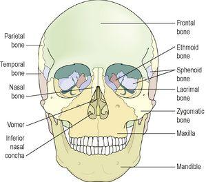
The skeleton of the face is formed by 13 bones in addition to the frontal bone already described.
Figure 16.16
shows the relationships between the bones:
•
2 zygomatic (cheek) bones
•
1 maxilla
•
2 nasal bones
•
2 lacrimal bones
•
1 vomer
•
2 palatine bones
•
2 inferior conchae
•
1 mandible.
Figure 16.16
The bones of the face
. Anterior view.
Zygomatic (cheek) bones
The zygomatic bone originates as two bones that fuse before birth. They form the prominences of the cheeks and part of the floor and lateral walls of the orbital cavities.
Maxilla (upper jaw bone)
This originates as two bones, but fusion takes place before birth. The maxilla forms the upper jaw, the anterior part of the roof of the mouth, the lateral walls of the nasal cavity and part of the floor of the orbital cavities. The
alveolar ridge
, or
process
, projects downwards and carries the upper teeth. On each side is a large air sinus, the
maxillary sinus
, lined with ciliated mucous membrane and with openings into the nasal cavity.
Nasal bones
These are two small flat bones that form the greater part of the lateral and superior surfaces of the bridge of the nose.
Lacrimal bones
These two small bones are posterior and lateral to the nasal bones and form part of the medial walls of the orbital cavities. Each is pierced by a foramen for the passage of the
nasolacrimal duct
that carries the tears from the medial canthus of the eye to the nasal cavity.
Vomer
The vomer is a thin flat bone that extends upwards from the middle of the hard palate to form most of the inferior part of the nasal septum. Superiorly it articulates with the perpendicular plate of the ethmoid bone.
Palatine bones
These are two small L-shaped bones. The horizontal parts unite to form the posterior part of the hard palate and the perpendicular parts project upwards to form part of the lateral walls of the nasal cavity. At their upper extremities they form part of the orbital cavities.
