Ross & Wilson Anatomy and Physiology in Health and Illness (178 page)
Read Ross & Wilson Anatomy and Physiology in Health and Illness Online
Authors: Anne Waugh,Allison Grant
Tags: #Medical, #Nursing, #General, #Anatomy

Bone structure
General structure of a long bone
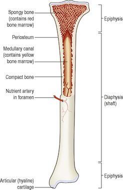
These have a diaphysis or shaft and two epiphyses or extremities (
Fig. 16.1
). The diaphysis is composed of compact bone with a central medullary canal, containing fatty yellow bone marrow. The epiphyses consist of an outer covering of compact bone with
spongy
(
cancellous
)
bone
inside. The diaphysis and epiphyses are separated by
epiphyseal cartilages
, which ossify when growth is complete. Thickening of a bone occurs by the deposition of new bone tissue under the periosteum.
Figure 16.1
A mature long bone
– partially sectioned.
Long bones are almost completely covered by a vascular membrane, the
periosteum
, which has two layers. The outer layer is tough and fibrous, and protects the bone underneath. The inner layer contains osteoblasts and osteoclasts, the cells responsible for bone production and breakdown (see below), and is important in repair and remodelling of the bone. The periosteum covers the whole bone except within joint cavities, allows attachments of tendons and is continuous with the joint capsule.
Hyaline cartilage
replaces periosteum on bone surfaces that form joints.
Blood and nerve supply
Blood supply to the shaft of the bone derives from one or more nutrient arteries; the epiphyses have their own blood supply, although in the mature bone the capillary networks arising from the two are heavily interconnected. The sensory supply usually enters the bone at the same site as the nutrient artery, and branches extensively throughout the bone. Bone injury is, therefore, usually very painful.
Structure of short, irregular, flat and sesamoid bones
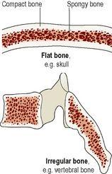
These have a relatively thin outer layer of compact bone with spongy bone inside containing red bone marrow (
Fig. 16.2
). They are enclosed by periosteum except the inner layer of the cranial bones where it is replaced by dura mater.
Figure 16.2
Sections of flat and irregular bones.
Microscopic structure of bone
Bone is a strong and durable type of connective tissue. Its major constituent (65%) is a mixture of calcium salts, mainly calcium phosphate. This inorganic matrix gives bone great hardness, but on its own would be brittle and prone to shattering. The remaining third is organic material, called
osteoid
, which is composed mainly of collagen. Collagen is very strong and gives bone slight flexibility. The cellular component of bone contributes less than 2% of bone mass.
Bone cells
The cells responsible for bone formation are
osteoblasts
(these later mature into
osteocytes
). Osteoblasts and
chondrocytes
(cartilage-forming cells) develop from the same parent fibrous tissue cells. Osteoclasts break down bone. They are large, multinucleate cells made from the fusion of up to 20 monocytes (
p. 63
).
A fine balance of osteoblast and osteoclast activity maintains normal bone structure and functions.
Osteoblasts
These bone-forming cells secrete both the organic and inorganic components of bone. They are present:
•
in the deeper layers of periosteum
•
in the centres of ossification of immature bone
•
at the ends of the diaphysis adjacent to the epiphyseal cartilages of long bones
•
at the site of a fracture.
Osteocytes
As bone develops, osteoblasts become trapped within the newly formed bone. They stop forming new bone at this stage and are called osteocytes. These are the mature bone cells that monitor and maintain bone tissue, and are nourished by tissue fluid in the canaliculi that radiate from the central canals.
Osteoclasts
Their function is resorption of bone to maintain the optimum shape. This takes place at bone surfaces:
•
under the periosteum, to maintain the shape of bones during growth and to remove excess callus formed during healing of fractures (
p. 384
)
•
round the walls of the medullary canal during growth and to canalise callus during healing.
Compact (cortical) bone
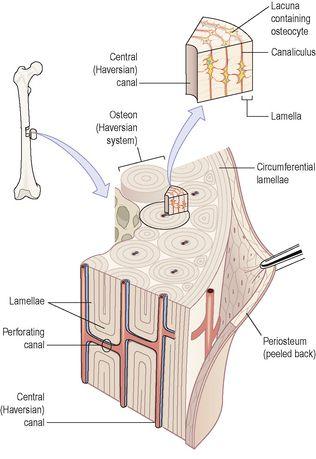
Compact bone makes up about 80% of the body bone mass. It is made up of a large number of parallel tube-shaped units called
osteons
(Haversian systems), each of which is made up of a central canal surrounded by a series of expanding rings, similar to the growth rings of a tree (
Fig. 16.3
). Osteons tend to be aligned the same way that force is applied to the bone, so for example in the femur (thigh bone), they run from one epiphysis to the other. This gives the bone great strength.
Figure 16.3
Microscopic structure of compact bone.
The central canal contains nerves, lymphatics and blood vessels, and each central canal is linked with neighbouring canals by tunnels running at right angles between them, called
perforating canals
. The series of cylindrical plates of bone arranged around each central canal are called
lamellae
. Between the adjacent lamellae of the osteon are strings of little cavities called
lacunae
, in each of which sits an osteocyte. Lacunae communicate with each other through a series of tiny channels called
canaliculi
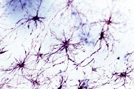
, which allows the circulation of interstitial fluid through the bone, and direct contact between the osteocytes, which extend fine processes into them (
Fig. 16.4
).
Figure 16.4
Light micrograph of osteocytes, showing the multiple fine processes that extend through bone canaliculi and allow each cell to communicate directly with its neighbours.
Between the osteons are
interstitial lamellae
, the remnants of older systems partially broken down during remodelling or growth of bone.
Spongy (cancellous, trabecular) bone
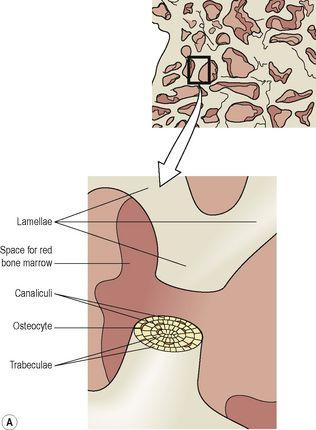
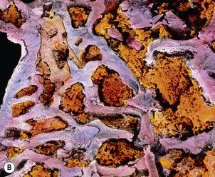
To the naked eye, spongy bone looks like a honeycomb. Microscopic examination reveals a framework formed from trabeculae (meaning ‘little beams’), which consist of a few lamellae and osteocytes interconnected by canaliculi (
Fig. 16.5
). Osteocytes are nourished by interstitial fluid seeping into the bone through the tiny canaliculi. The spaces between the trabeculae contain red bone marrow. In addition, spongy bone is lighter than compact bone, reducing the weight of the skeleton.
Figure 16.5
A.
Microscopic structure of spongy bone.
B.
Electron micrograph of spongy bone showing bone marrow (orange) filling the spaces between trabeculae (grey/blue).
Development of bone tissue
