Ross & Wilson Anatomy and Physiology in Health and Illness (87 page)
Read Ross & Wilson Anatomy and Physiology in Health and Illness Online
Authors: Anne Waugh,Allison Grant
Tags: #Medical, #Nursing, #General, #Anatomy

The semicircular canals have no auditory function although they are closely associated with the cochlea. Instead they provide information about the position of the head in space, contributing to maintenance of posture and balance.
There are three semicircular canals, one lying in each of the three planes of space. They are situated above, beside and behind the vestibule of the inner ear and open into it.
The semicircular canals, like the cochlea, are composed of an outer bony wall and inner membranous tubes or
ducts
. The membranous ducts contain endolymph and are separated from the bony wall by perilymph.
The utricle
is a membranous sac which is part of the vestibule and the three membranous ducts open into it at their dilated ends, the
ampullae
. The saccule is a part of the vestibule and communicates with the utricle and the cochlea.
In the walls of the utricle, saccule and ampullae are fine, specialised epithelial cells with minute projections, called hair cells. Amongst the
hair cells
there are receptors on sensory nerve endings, which combine forming the vestibulocochlear nerve.
Physiology of balance
The semicircular canals and the vestibule (utricle and saccule) are concerned with balance. Any change of position of the head causes movement in the perilymph and endolymph, which bends the hair cells and stimulates the sensory receptors in the utricle, saccule and ampullae. The resultant nerve impulses are transmitted by the vestibular nerve, which joins the cochlear nerve to form the vestibulocochlear nerve. The vestibular branch passes first to the
vestibular nucleus
, then to the cerebellum.
The cerebellum also receives nerve impulses from the eyes and proprioceptors (sensory receptors) in the skeletal muscles and joints. Impulses from these three sources are coordinated and efferent nerve impulses pass to the cerebrum and to skeletal muscles. This results in awareness of body position, maintenance of upright posture and fixing of the eyes on the same point, independently of head movements.
Sight and the eye
Learning outcomes
After studying this section, you should be able to:
describe the gross structure of the eye
describe the route taken by nerve impulses from the retina to the cerebrum
explain how light entering the eye is focused on the retina
state the functions of the extraocular eye muscles
explain the functions of the accessory organs of the eye.
The eye is the organ of sight. It is situated in the orbital cavity and supplied by the
optic nerve
(2nd cranial nerve).
It is almost spherical in shape and about 2.5 cm in diameter. The space between the eye and the orbital cavity is occupied by adipose tissue. The bony walls of the orbit and the fat help to protect the eye from injury.
Structurally the two eyes are separate but, unlike the ear, some of their activities are coordinated so that they function as a pair. It is possible to see with only one eye (monocular vision), but three-dimensional vision is impaired when only one eye is used, especially in relation to the judgement of speed and distance.
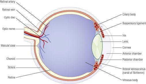
Structure (
Fig. 8.8
)
There are three layers of tissue in the walls of the eye:
•
the outer fibrous layer: sclera and cornea
•
the middle vascular layer or
uveal tract
: consisting of the choroid, ciliary body and iris
•
the inner nervous tissue layer: retina.
Figure 8.8
Section of the eye.
Structures inside the eyeball include the lens, aqueous fluid and vitreous body.
Sclera and cornea
The sclera, or white of the eye, forms the outermost layer of the posterior and lateral aspects of the eyeball and is continuous anteriorly with the transparent cornea. It consists of a firm fibrous membrane that maintains the shape of the eye and gives attachment to the
extrinsic muscles
of the eye (
p. 198
).
Anteriorly the sclera continues as a clear transparent epithelial membrane, the cornea. Light rays pass through the cornea to reach the retina. The cornea is convex anteriorly and is involved in refracting (bending) light rays to focus them on the retina.
The choroid lines the posterior five-sixths of the inner surface of the sclera. It is very rich in blood vessels and is deep chocolate brown in colour. Light enters the eye through the pupil, stimulates the sensory receptors in the retina and is then absorbed by the choroid.
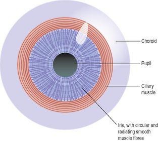
Figure 8.9
The choroid, ciliary body and iris.
Viewed from the front.


