Ross & Wilson Anatomy and Physiology in Health and Illness (76 page)
Read Ross & Wilson Anatomy and Physiology in Health and Illness Online
Authors: Anne Waugh,Allison Grant
Tags: #Medical, #Nursing, #General, #Anatomy

I.
Olfactory: sensory
II.
Optic: sensory
III.
Oculomotor: motor
IV.
Trochlear: motor
V.
Trigeminal: mixed
VI.
Abducent: motor
VII.
Facial: mixed
VIII.
Vestibulocochlear (auditory): sensory
IX.
Glossopharyngeal: mixed
X.
Vagus: mixed
XI.
Accessory: motor
XII.
Hypoglossal: motor.
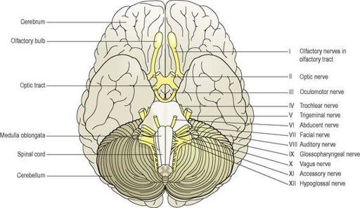
Figure 7.43
The inferior surface of the brain showing the cranial nerves and associated structures.
I Olfactory nerves (sensory)
These are the nerves of the
sense of smell
. Their sensory receptors and fibres originate in the upper part of the mucous membrane of the nasal cavity, pass upwards through the cribriform plate of the ethmoid bone and then pass to the
olfactory bulb
(see
Fig. 8.24
,
p. 200
). The nerves then proceed backwards as the olfactory tract, to the area for the perception of smell in the temporal lobe of the cerebrum (
Ch. 8
).
II Optic nerves (sensory)
These are the nerves of the
sense of sight
. The fibres originate in the retinae of the eyes and they combine to form the optic nerves (see
Fig. 8.13
,
p. 194
). They are directed backwards and medially through the posterior part of the orbital cavity. They then pass through the
optic foramina
of the sphenoid bone into the cranial cavity and join at the
optic chiasma
. The nerves proceed backwards as the
optic tracts
to the
lateral geniculate bodies
of the thalamus. Impulses pass from there to the visual areas in the occipital lobes of the cerebrum and to the cerebellum. In the occipital lobe sight is perceived, and in the cerebellum the impulses from the eyes contribute to the maintenance of balance, posture and orientation of the head in space.
III Oculomotor nerves (motor)
These nerves arise from nuclei near the cerebral aqueduct. They supply:
•
four of the six extrinsic muscles, which move the eyeball, i.e. the
superior
,
medial
and
inferior recti
and the
inferior oblique muscle
(see
Table 8.1
,
p. 198
)
•
the intrinsic (intraocular) muscles:
–
ciliary muscles
, which alter the shape of the lens, changing its refractive power
–
circular muscles
of the iris, which constrict the pupil
•
the
levator palpebrae muscles
, which raise the upper eyelids.
IV Trochlear nerves (motor)
These nerves arise from nuclei near the cerebral aqueduct. They supply the
superior oblique muscles
of the eyes.
V Trigeminal nerves (mixed)
These nerves contain motor and sensory fibres and are among the largest of the cranial nerves. They are the chief sensory nerves for the face and head (including the oral and nasal cavities and teeth), receiving impulses of pain, temperature and touch. The motor fibres stimulate the muscles of mastication.
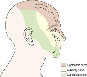
As the name suggests, there are three main branches of the trigeminal nerves. The dermatomes innervated by the sensory fibres on the right side are shown in
Figure 7.44
.
Figure 7.44
The cutaneous distribution of the main branches of the right trigeminal nerve.
The ophthalmic nerves
are sensory only and supply the lacrimal glands, conjunctiva of the eyes, forehead, eyelids, anterior aspect of the scalp and mucous membrane of the nose.
The maxillary nerves
are sensory only and supply the cheeks, upper gums, upper teeth and lower eyelids.
The mandibular nerves
contain both sensory and motor fibres. These are the largest of the three divisions and they supply the teeth and gums of the lower jaw, pinnae of the ears, lower lip and tongue. The motor fibres supply the muscles of mastication (chewing).
VI Abducent nerves (motor)
These nerves arise from nuclei lying under the floor of the fourth ventricle. They supply the
lateral rectus muscles
of the eyeballs causing abduction, as the name suggests.
VII Facial nerves (mixed)
These nerves are composed of both motor and sensory nerve fibres, arising from nuclei in the lower part of the pons. The motor fibres supply the muscles of facial expression. The sensory fibres convey impulses from the taste buds in the anterior two-thirds of the tongue to the taste perception area in the cerebral cortex (see
Fig. 7.21
).
VIII Vestibulocochlear (auditory) nerves (sensory)
These nerves are composed of two divisions, the vestibular nerves and cochlear nerves.
The vestibular nerves
arise from the semicircular canals of the inner ear and convey impulses to the cerebellum. They are associated with the maintenance of posture and balance.
The cochlear nerves
originate in the spiral organ (of Corti) in the inner ear and convey impulses to the hearing areas in the cerebral cortex where sound is perceived.
IX Glossopharyngeal nerves (mixed)
The motor fibres arise from nuclei in the medulla oblongata and stimulate the muscles of the tongue and pharynx and the secretory cells of the parotid (salivary) glands.
The sensory fibres convey impulses to the cerebral cortex from the posterior third of the tongue, the tonsils and pharynx and from taste buds in the tongue and pharynx. These nerves are essential for the swallowing and gag reflexes. Some fibres conduct impulses from the carotid sinus, which plays an important role in the control of blood pressure (
p. 88
).
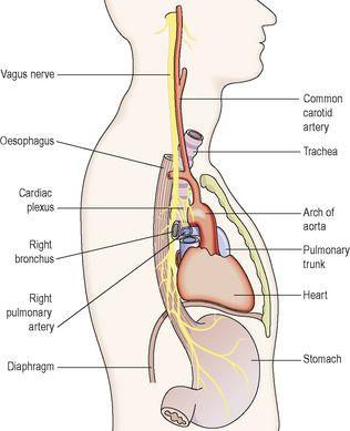
X Vagus nerves (mixed) (
Fig. 7.45
)
These nerves have a more extensive distribution than any other cranial nerves and their name aptly means ‘wanderer’. They pass down through the neck into the thorax and the abdomen. These nerves form an important part of the parasympathetic nervous system (see
Fig. 7.47
).
Figure 7.45
The position of the vagus nerve in the thorax viewed from the right side.
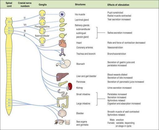
Figure 7.47
The parasympathetic outflow, the main structures supplied and the effects of stimulation.
Solid blue lines – preganglionic fibres; broken lines – postganglionic fibres. Where there are no broken lines, the postganglionic neurone is in the wall of the structure.
The motor fibres arise from nuclei in the medulla and supply the smooth muscle and secretory glands of the pharynx, larynx, trachea, bronchi, heart, carotid body, oesophagus, stomach, intestines, exocrine pancreas, gall bladder, bile ducts, spleen, kidneys, ureter and blood vessels in the thoracic and abdominal cavities.
The sensory fibres convey impulses from the membranes lining the same structures to the brain.
XI Accessory nerves (motor)
These nerves arise from nuclei in the medulla oblongata and in the spinal cord. The fibres supply the
sternocleidomastoid
and
trapezius muscles
. Branches join the vagus nerves and supply the
pharyngeal
and
laryngeal muscles
.
XII Hypoglossal nerves (motor)
These nerves arise from nuclei in the medulla oblongata. They supply the muscles of the tongue and muscles surrounding the hyoid bone and contribute to swallowing and speech.
Autonomic nervous system


