Stretching Anatomy-2nd Edition (2 page)
Read Stretching Anatomy-2nd Edition Online
Authors: Arnold Nelson,Jouko Kokkonen
Tags: #Science, #Life Sciences, #Human Anatomy & Physiology

The initial length of a sarcomere is an important factor in muscle function. The amount of force produced by each sarcomere is influenced by length in a pattern similar in shape to an upside-down letter U. As such, force is reduced when the sarcomere length is either long or short. As the sarcomere lengthens, only the tips of the thick and thin filaments can contact each other, and this reduces the number of force-producing connections between the two filaments. When the sarcomere shortens, the thin filaments start to overlap each other, and this overlap also reduces the number of positive force-producing connections.
Sarcomere length is controlled by proprioceptors, or specialized structures incorporated within the muscle organs, especially within the muscles of the limbs. The proprioceptors are specialized sensors that provide information about joint angle, muscle length, and muscle tension. Information about changes in muscle length is provided by proprioceptors called muscle spindles, and they lie parallel to the muscle cells. The Golgi tendon organs, or GTOs, the other type of proprioceptor, lie in series with the muscle cells. GTOs provide information about changes in muscle tension and indirectly can influence muscle length. The muscle spindle has a fast dynamic component and a slow static component that provides information on the amount and rate of change in length. Fast length changes can trigger a stretch, or myotatic, reflex that attempts to resist the change in muscle length by causing the stretched muscle to contract. Slower stretches allow the muscle spindles to relax and adapt to the new longer length.
When the muscle contracts it produces tension in the tendon and the GTOs. The GTOs record the change and rate of change in tension. When this tension exceeds a certain threshold, it triggers the lengthening reaction via spinal cord connections to inhibit the muscles from contracting and cause them to relax. Also, muscle contraction can induce reciprocal inhibition, or the relaxation of the opposing muscles. For instance, a hard contraction of the biceps brachii can induce relaxation within the triceps brachii.
The body adapts differently to acute stretching (or short-term stretching) and chronic stretching (or stretching done multiple times during a week). The majority of current research shows that when acute stretches cause a noticeable increase in a joint’s range of motion, the person can experience either inhibition of the motor nerves, overlengthening of the muscle sarcomeres, or increased length and compliance of the muscle’s tendons. No one is sure of the extent of these changes, but it appears that the muscle shape and cell arrangement, muscle length and contribution to movement, and length of the distal and proximal tendons all play a role. Nevertheless, these transient changes are manifested as decreases in maximal strength, power, and strength endurance. On the other hand, research studies have shown that regular heavy stretching for a minimum of 10 to 15 minutes three or four days a week (chronic stretching) results in the development of increased strength, power, and strength endurance as well as improved flexibility and mobility. Animal studies suggest that these benefits are due in part to increased numbers of sarcomeres in series.
Likewise, research into stretching for injury prevention has shown differences between acute stretching and chronic stretching. Although acute stretching can help an extremely tight person reduce the incidence of muscle strains, the normal person appears to gain minimal injury-prevention benefit from acute stretching. People who are inherently more flexible are less prone to exercise-related injuries, and inherent flexibility is increased with heavy stretching three or four days a week. Because of these differences between acute and chronic stretching, many exercise experts now encourage people to do the majority of their stretching at the end of a workout.
Types of Stretches
The stretches featured in this book can be executed in a variety of ways. Most people prefer to do these stretches on their own, but they can also be done with the help of another person. Stretches performed without assistance are referred to as active stretches. Stretches performed with assistance from another person are called passive stretches.
There are four major types of stretches: static, ballistic, proprioceptive neuromuscular facilitation (PNF), and dynamic.
The
static stretch
is the most common. In static stretching, you stretch a particular muscle or group of muscles by holding that stretch for a period of time.
Ballistic stretches
involve bouncing movements and do not involve holding the stretch for any length of time. Since ballistic stretching can activate the stretch reflex, many people have postulated that ballistic stretching has a greater potential to cause muscle or tendon damage, especially in the tightest muscles. However, this assertion is purely speculative, and no published research supports the claim that ballistic stretching can cause injury.
Proprioceptive neuromuscular facilitation (PNF) stretching
refers to a stretching technique that tries to more fully incorporate the actions of the proprioceptors by stretching a contracted muscle through the joint’s range of motion. After moving through the complete range of motion, the muscle is relaxed and rested before it is stretched again. This type of stretching is best done with the assistance of another person.
Dynamic stretching
is a more functionally oriented stretch that uses sport-specific movements to move the limbs through a greater range of motion than normal. Dynamic stretching is generally characterized by swinging, jumping, or exaggerated movements in which the momentum of the movement carries the limbs to or past the regular limits of the range of motion and activates a proprioceptive reflex response. The proper activation of the proprioceptors can cause facilitation of the nerves that activated the muscle cells. This facilitation enables the nerves to fire more quickly, thus enabling the muscle to make fast and more powerful contractions. Since dynamic stretches increase both muscle temperature and proprioceptive activation, dynamic stretching has been found to be advantageous for improving athletic performance. Dynamic stretching should not be confused with ballistic stretching. Although both involve repeated movements, ballistic movements are rapid, bouncing movements that involve small ranges of movement near the end of the range of motion.
Benefits of a Stretching Program
Several chronic training benefits can be gained through a regular stretching program (see chapter 9 for specific programs):
- Improved flexibility, stamina (muscular endurance), and muscular strength (the degree of benefit depends on how much stress is put on the muscle; chapter 9 explains how this should be done)
- Reduced muscle soreness
- Improved muscle and joint mobility
- More efficient muscular movements and fluidity of motion
- Greater ability to exert maximum force through a wider range of motion
- Prevention of some lower-back problems
- Improved appearance and self-image
- Improved body alignment and posture
- Better warm-up and cool-down in an exercise session
- Improved maintenance of blood glucose
Static and Dynamic Stretches for Athletes
Many athletes use static and dynamic stretches in their training programs. Static stretches improve flexibility in certain muscle–joint areas. This type of stretching is the most common approach for improving flexibility. In static stretching, you hold a stretch of a particular muscle or muscle group for a period of time.
Some athletes prefer using dynamic stretches, particularly as a part of a warm-up or as a preparation for competition. Dynamic stretches stimulate the proprioceptors (stretch receptors), activating their response in an aggressive way by sending feedback to the stretched muscles to be contracted after a quick bouncing motion. Because some athletic events, such as explosive, short-duration activities, could possibly enhance the stimulation of this proprioceptive activation, dynamic stretching prepares athletes better for explosive movements. Such explosive movements might be required to accomplish a certain goal in an athletic event. For example, a person can jump farther and higher if he does a couple of quick up and down movements, flexing and extending the hips and knees.
How to Use This Book
The first seven chapters of this book highlight stretches for the major joint areas of the body, beginning with the neck and ending with the feet and calves. Within each chapter are several stretches targeting the muscles involved in moving the joints in each part of the body. The movements targeting what are likely to be the stiffest muscles include a progression of stretches so that the person with the tightest muscles (beginner) is not trying to do a stretch that puts too much stress on the joint and results in muscle, ligament, and tendon damage. As you increase in flexibility, graduate to the next level.
Chapter 8 contains nine dynamic stretches that encompass all the major joint areas. Chapter 9 contains suggested stretching programs for beginners through advanced as well as a program shown to lower blood glucose. In addition, chapter 9 includes sport-specific stretching routines. If you are interested in a specific sport, these tables will guide you to the stretches to use in your training to ensure that you target the most important muscle groups used in that sport.
The name of each stretch indicates the major movements of the muscles being stretched. As such, you should remember that to stretch a specific muscle, the stretch must involve one or more movements in the opposite direction of the desired muscle’s movements. The illustrations depict the body positions used for each stretch as well as the muscles being stretched. The muscles most stretched are illustrated in a dark red (see key), and any nearby muscles that are less stretched are illustrated in a lighter red.

In addition to the illustrations, each stretch contains three sections:
- Execution, which provides step-by-step instructions on how to perform the stretch
- Muscles stretched, which provides the names of the muscles being stretched
- Stretch notes, which provide specific information concerning the how and why behind the need for the stretch as well as any safety considerations
Chapter 1
Neck
The seven cervical vertebrae along with associated muscles and ligaments make up the flexible framework of the neck. The vertebrae, muscles, and ligaments work together to support and move the head. The first and second cervical vertebrae have unique shapes and are called the
atlas
and
axis
. The atlas is a bony ring that supports the skull. The axis has an upward peglike projection, the dens, that gives the atlas a point to pivot around. The axis and the other five cervical vertebrae have a posterior bony protuberance, or spinous process, that attaches to the large, thick nuchal ligament. The vertebral bodies (the oval-shaped bone mass) are connected by posterior and anterior ligaments, along with other ligaments that connect each spinous and transverse (lateral bony protuberance) process to their corresponding parts on the adjacent vertebrae. In addition, each vertebra is separated by an intervertebral disc. Through compression of the vertebrae upon the discs, the neck can move forward, backward, and sideways.
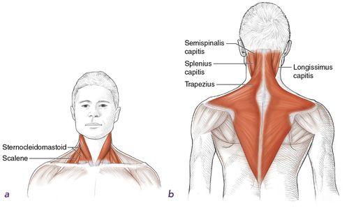
The neck muscles are located in two triangular regions called the anterior (front) and posterior (back) triangles. The borders of the anterior triangle are the mandible (jawbone), the sternum (breastbone), and the sternocleidomastoid muscle. The major anterior muscles are the sternocleidomastoid and scalene (
figure 1.1
a
). The borders of the posterior triangle are the clavicle (collarbone), sternocleidomastoid muscle, and trapezius muscle. The major posterior muscles (
figure 1.1
b
) are the trapezius, longissimus capitis, semispinalis capitis, and splenius capitis.
Figure 1.1
Neck muscles:
(a)
anterior;
(b)
posterior.