Ross & Wilson Anatomy and Physiology in Health and Illness (109 page)
Read Ross & Wilson Anatomy and Physiology in Health and Illness Online
Authors: Anne Waugh,Allison Grant
Tags: #Medical, #Nursing, #General, #Anatomy

During swallowing, the nasal and oral parts are separated by the soft palate and the
uvula
.
The laryngopharynx
The laryngeal part of the pharynx extends from the oropharynx above and continues as the oesophagus below, i.e. from the level of the 3rd to the 6th cervical vertebrae.
Structure
The walls of the pharynx contain several types of tissue.
Mucous membrane lining
The mucosa varies slightly in the different regions. In the nasopharynx it is continuous with the lining of the nose and consists of ciliated columnar epithelium; in the oropharynx and laryngopharynx it is formed by tougher stratified squamous epithelium, which is continuous with the lining of the mouth and oesophagus. This lining protects underlying tissues from the abrasive action of foodstuffs passing through during swallowing.
Submucosa
The layer of tissue below the epithelium (the submucosa) is rich in mucosa-associated lymphoid tissue (
MALT, p. 133
), involved in protection against infection. Tonsils are masses of MALT that bulge through the epithelium. Some glandular tissue is also found here.
Smooth muscle
The pharyngeal muscles help to keep the pharynx permanently open so that breathing is not interfered with. Sometimes in sleep, and particularly if sedative drugs or alcohol have been taken, the tone of these muscles is reduced and the opening through the pharynx can become partially or totally obstructed. This contributes to snoring and periodic wakenings, which disturb sleep.
Constrictor muscles are responsible for constricting the pharynx during swallowing, pushing food and fluid into the oesophagus.
Blood and nerve supply
Blood is supplied to the pharynx by several branches of the facial artery. The venous return is into the facial and internal jugular veins.
The nerve supply is from the pharyngeal plexus, formed by parasympathetic and sympathetic nerves. Parasympathetic supply is by the
vagus
and
glossopharyngeal
nerves. Sympathetic supply is by nerves from the
superior cervical ganglia
(
p. 168
).
Functions
Passageway for air and food
The pharynx is involved in both the respiratory and the digestive systems: air passes through the nasal and oral sections, and food through the oral and laryngeal sections.
Warming and humidifying
By the same methods as in the nose, the air is further warmed and moistened as it passes through the pharynx.
Taste
There are olfactory nerve endings of the sense of taste in the epithelium of the oral and pharyngeal parts.
Hearing
The auditory tube, extending from the nasopharynx to each middle ear, allows air to enter the middle ear. Satisfactory hearing depends on the presence of air at atmospheric pressure on each side of the
tympanic membrane
(eardrum,
p. 187
).
Protection
The lymphatic tissue of the pharyngeal and laryngeal tonsils produces antibodies in response to antigens, e.g. bacteria (
Ch. 15
). The tonsils are larger in children and tend to atrophy in adults.
Speech
The pharynx functions in speech; by acting as a resonating chamber for sound ascending from the larynx, it helps (together with the sinuses) to give the voice its individual characteristics.
Larynx
Learning outcomes
After studying this section, you should be able to:
describe the structure and function of the larynx
outline the physiology of speech generation.
Position
The larynx or ‘voice box’ links the laryngopharynx and the trachea. It lies at the level of the 3rd, 4th, 5th and 6th cervical vertebrae. Until puberty there is little difference in the size of the larynx between the sexes. Thereafter, it grows larger in the male, which explains the prominence of the ‘Adam’s apple’ and the generally deeper voice.
Structures associated with the larynx
Superiorly
– the hyoid bone and the root of the tongue
Inferiorly
– it is continuous with the trachea
Anteriorly
– the muscles attached to the hyoid bone and the muscles of the neck
Posteriorly
– the laryngopharynx and 3rd to 6th cervical vertebrae
Laterally
– the lobes of the thyroid gland.
Structure
Cartilages
The larynx is composed of several irregularly shaped cartilages attached to each other by ligaments and membranes. The main cartilages are:
|
| |
| • 1 epiglottis. | elastic fibrocartilage |
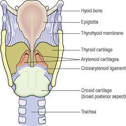
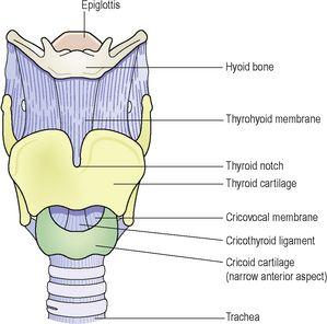
The thyroid cartilage
(
Figs 10.5
and
10.6
). This is the most prominent of the laryngeal cartilages. Made of hyaline cartilage, it lies to the front of the neck. Its anterior wall projects into the soft tissues of the front of the throat, forming the
laryngeal prominence
or Adam’s apple, which is easily felt and often visible in adult males. The anterior wall is partially divided by the
thyroid notch
. The cartilage is incomplete posteriorly, and is bound with ligaments to the hyoid bone above and the cricoid cartilage below.
Figure 10.5
Larynx
– viewed from behind.
Figure 10.6
Larynx
– viewed from the front.
The upper part of the thyroid cartilage is lined with stratified squamous epithelium like the larynx, and the lower part with ciliated columnar epithelium like the trachea. There are many muscles attached to its outer surface.
The thyroid cartilage forms most of the anterior and lateral walls of the larynx.
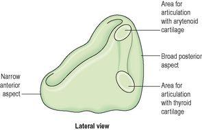
The cricoid cartilage (
Fig. 10.7
)
This lies below the thyroid cartilage and is also composed of hyaline cartilage. It is shaped like a signet ring, completely encircling the larynx with the narrow part anteriorly and the broad part posteriorly. The broad posterior part articulates with the arytenoid cartilages and with the thyroid cartilage. It is lined with ciliated columnar epithelium and there are muscles and ligaments attached to its outer surface (
Fig. 10.7
). The lower border of the cricoid cartilage marks the end of the upper respiratory tract.


