Ross & Wilson Anatomy and Physiology in Health and Illness (132 page)
Read Ross & Wilson Anatomy and Physiology in Health and Illness Online
Authors: Anne Waugh,Allison Grant
Tags: #Medical, #Nursing, #General, #Anatomy

outline the arrangement of normal primary and secondary dentition.

The mouth or oral cavity is bounded by muscles and bones:
Anteriorly
– by the lips
Posteriorly
– it is continuous with the oropharynx
Laterally
– by the muscles of the cheeks
Superiorly
– by the bony hard palate and muscular soft palate
Inferiorly
– by the muscular tongue and the soft tissues of the floor of the mouth.
The oral cavity is lined throughout with
mucous membrane
, consisting of stratified squamous epithelium containing small mucus-secreting glands.
The part of the mouth between the gums and the cheeks is the
vestibule
and the remainder of the cavity is the
oral cavity
. The mucous membrane lining of the cheeks and the lips is reflected onto the gums or
alveolar ridges
and is continuous with the skin of the face.
The
palate
forms the roof of the mouth and is divided into the anterior
hard palate
and the posterior
soft palate
(
Fig. 12.1
). The hard palate is formed by the maxilla and the palatine bones. The soft palate is muscular, curves downwards from the posterior end of the hard palate and blends with the walls of the pharynx at the sides.
The
uvula
is a curved fold of muscle covered with mucous membrane, hanging down from the middle of the free border of the soft palate. Originating from the upper end of the uvula are four folds of mucous membrane, two passing downwards at each side to form membranous arches. The posterior folds, one on each side, are the
palatopharyngeal
arches and the two anterior folds are the
palatoglossal arches
. On each side, between the arches, is a collection of lymphoid tissue called the
palatine tonsil
.
Tongue
The tongue is a voluntary muscular structure that occupies the floor of the mouth. It is attached by its base to the
hyoid bone
(see
Fig. 10.4
,
p. 236
) and by a fold of its mucous membrane covering, called the
frenulum
, to the floor of the mouth (
Fig. 12.8
). The superior surface consists of stratified squamous epithelium, with numerous
papillae
(little projections). Many of these contain sensory receptors (specialised nerve endings) for the sense of taste in the
taste buds
(see
Fig. 8.25, p. 201
). There are several types of papilla; however no clear relationship between these and discrimination of different tastes has been found.
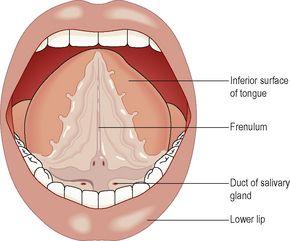
Figure 12.8
The inferior surface of the tongue.
Blood supply
The main arterial blood supply to the tongue is by the
lingual branch
of the
external carotid artery
. Venous drainage is by the
lingual vein
, which joins the
internal jugular vein
.
Nerve supply
The nerves involved are:
•
the
hypoglossal nerves
(12th cranial nerves), which supply the voluntary muscle
•
the
lingual branch of the mandibular nerves
, which arise from the 5th cranial nerves, are the nerves of somatic (ordinary) sensation, i.e. pain, temperature and touch
•
the
facial
and
glossopharyngeal nerves
(7th and 9th cranial nerves), the nerves of taste.
Functions of the tongue
The tongue plays an important part in:
•
chewing (mastication)
•
swallowing (deglutition)
•
speech (
p. 240
)
•
taste (
p. 200
).
Nerve endings of the sense of taste are present in the papillae and widely distributed in the epithelium of the tongue, soft palate, pharynx and epiglottis.
Teeth
The teeth are embedded in the alveoli or sockets of the alveolar ridges of the mandible and the maxilla (
Fig. 12.9
). Babies are born with two sets, or
dentitions
, the
temporary
or
deciduous teeth
and the
permanent teeth
(
Fig. 12.10
). At birth the teeth of both dentitions are present, in immature form, in the mandible and maxilla.
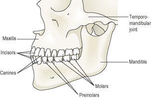
Figure 12.9
The permanent teeth and the jaw bones.
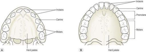
Figure 12.10
The roof of the mouth. A.
The deciduous teeth – viewed from below.
B.
The permanent teeth – viewed from below.
There are 20 temporary teeth, 10 in each jaw. They begin to erupt when the child is about 6 months old, and should all be present by 24 months (
Table 12.1
).
Table 12.1
Deciduous and permanent dentitions
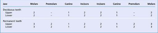
The permanent teeth begin to replace the deciduous teeth in the 6th year of age and this dentition, consisting of 32 teeth, is usually complete by the 21st year.
Functions of the teeth
Teeth have different shapes depending on their functions.
Incisors
and
canine
teeth are the cutting teeth and are used for biting off pieces of food, whereas the
premolar
and
molar
teeth, with broad, flat surfaces, are used for grinding or chewing food (
Fig. 12.11
).
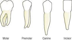
Figure 12.11
The shapes of the permanent teeth.
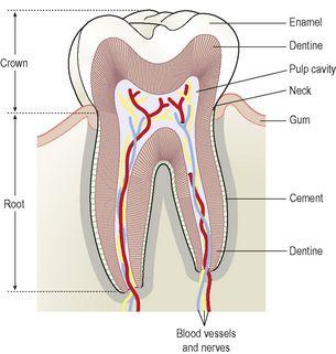
Structure of a tooth (
Fig. 12.12
)
Although the shapes of the different teeth vary, the structure is the same and consists of:
•
the crown
– the part that protrudes from the gum
•
the root
– the part embedded in the bone
•
the neck
– the slightly narrowed region where the crown merges with the root.