Ross & Wilson Anatomy and Physiology in Health and Illness (64 page)
Read Ross & Wilson Anatomy and Physiology in Health and Illness Online
Authors: Anne Waugh,Allison Grant
Tags: #Medical, #Nursing, #General, #Anatomy

Figure 7.1
Functional components of the nervous system.
In turn the motor division is involved in activities that are:
•
voluntary
– the somatic nervous system (movement of voluntary muscles)
•
involuntary
– the autonomic nervous system (functioning of smooth and cardiac muscle and glands). The autonomic nervous system has two divisions:
sympathetic
and
parasympathetic
.
The first sections of this chapter explore the structure and functions of the components of the nervous system, while the final one considers the effects of body function when normal structures do not function normally.
Cells and tissues of the nervous system
Learning outcomes
After studying this section you should be able to:
compare and contrast the structure and functions of myelinated and unmyelinated neurones
state the functions of sensory and motor nerves
explain the events that occur following release of a neurotransmitter at a synapse
briefly describe the functions of four types of neuroglial cells
outline the response of nervous tissue to injury.
The nervous system consists of
neurones
, which conduct nerve impulses and are supported by unique connective tissue cells known as
neuroglia
. There are vast numbers of cells, 1 trillion (10
12
) glial cells and ten times fewer (10
11
) neurones.
Neurones (
Fig. 7.2
)
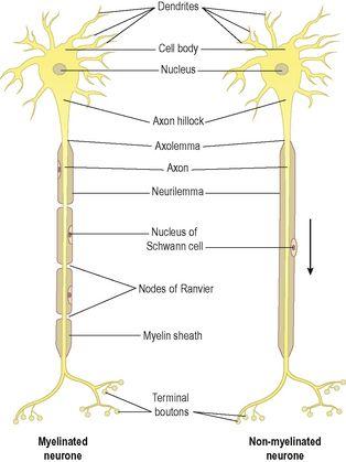
Each neurone (
Fig. 7.2
) consists of a
cell body
and its processes, one
axon
and many
dendrites
. Neurones are commonly referred to as nerve cells. Bundles of axons bound together are called
nerves
. Neurones cannot divide, and for survival they need a continuous supply of oxygen and glucose. Unlike many other cells, neurones can synthesise chemical energy (ATP) only from glucose.
Figure 7.2
The structure of neurones.
Arrow indicates direction of impulse conduction.
Neurones generate and transmit electrical impulses called
action potentials.
The initial strength of the impulse is maintained throughout the length of the neurone. Some neurones initiate nerve impulses while others act as ‘relay stations’ where impulses are passed on and sometimes redirected.
Nerve impulses can be initiated in response to stimuli from:
•
outside the body, e.g. touch, light waves
•
inside the body, e.g. a change in the concentration of carbon dioxide in the blood alters respiration; a thought may result in voluntary movement.
Transmission of nerve signals is both electrical and chemical. The action potential travelling down the nerve axon is an electrical signal, but because nerves do not come into direct contact with each other, the signal between a nerve cell and the next cell in the chain is chemical (
p. 141
).
Cell bodies
Nerve cells vary considerably in size and shape but they are all too small to be seen by the naked eye. Cell bodies form the
grey matter
of the nervous system and are found at the periphery of the brain and in the centre of the spinal cord. Groups of cell bodies are called
nuclei
in the central nervous system and
ganglia
in the peripheral nervous system. An important exception is the basal ganglia (nuclei) situated within the cerebrum (
p. 149
).
Axons and dendrites
Axons and dendrites are extensions of cell bodies and form the
white matter
of the nervous system. Axons are found deep in the brain and in groups, called
tracts
, at the periphery of the spinal cord. They are referred to as
nerves
or
nerve fibres
outside the brain and spinal cord.
Axons
Each nerve cell has only one axon, which begins at a tapered area of the cell body, the
axon hillock.
They carry impulses away from the cell body and are usually longer than the dendrites, sometimes as long as 100 cm.
Structure of an axon
The membrane of the axon is called the
axolemma
and it encloses the cytoplasmic extension of the cell body.
Myelinated neurones
Large axons and those of peripheral nerves are surrounded by a
myelin sheath
(
Fig. 7.3A
). This consists of a series of
Schwann cells
arranged along the length of the axon. Each one is wrapped around the axon so that it is covered by a number of concentric layers of Schwann cell plasma membrane. Between the layers of plasma membrane there is a small amount of fatty substance called
myelin
. The outermost layer of the Schwann cell plasma membrane is the
neurilemma
. There are tiny areas of exposed axolemma between adjacent Schwann cells, called
nodes of Ranvier
, which assist the rapid transmission of nerve impulses in myelinated neurones (
Fig. 7.2
).
Figure 7.4
shows a section through a nerve fibre at a node of Ranvier where the area without myelin can be clearly seen.


