Ross & Wilson Anatomy and Physiology in Health and Illness (60 page)
Read Ross & Wilson Anatomy and Physiology in Health and Illness Online
Authors: Anne Waugh,Allison Grant
Tags: #Medical, #Nursing, #General, #Anatomy

Lymph nodes are oval or bean-shaped organs that lie, often in groups, along the length of lymph vessels. The lymph drains through a number of nodes, usually 8 to 10, before returning to the venous circulation. These nodes vary considerably in size: some are as small as a pin head and the largest are about the size of an almond.
Structure
Lymph nodes (
Fig. 6.4
) have an outer capsule of fibrous tissue that dips down into the node substance forming partitions, or
trabeculae
. The main substance of the node consists of reticular and lymphatic tissue containing many lymphocytes and macrophages.
Figure 6.4
Section through a lymph node
. Arrows indicate the direction of lymph flow.
As many as four or five
afferent
lymph vessels may enter a lymph node while only one
efferent
vessel carries lymph away from the node. Each node has a concave surface called the
hilum
where an artery enters and a vein and the efferent lymph vessel leave.
The large numbers of lymph nodes situated in strategic positions throughout the body are arranged in deep and superficial groups.
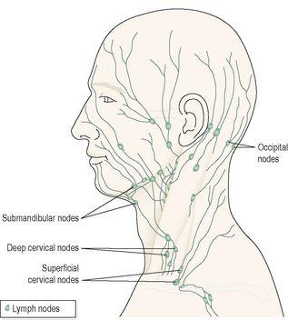
Lymph from the head and neck passes through deep and superficial
cervical nodes
(
Fig. 6.5
).
Figure 6.5
Some lymph nodes of the face and neck.
Lymph from the upper limbs passes through nodes situated in the elbow region, then through the deep and superficial
axillary nodes
.
Lymph from organs and tissues in the thoracic cavity drains through groups of nodes situated close to the mediastinum, large airways, oesophagus and chest wall. Most of the lymph from the breast passes through the axillary nodes.
Lymph from the pelvic and abdominal cavities passes through many lymph nodes before entering the cisterna chyli. The abdominal and pelvic nodes are situated mainly in association with the blood vessels supplying the organs and close to the main arteries, i.e. the aorta and the external and internal iliac arteries.
The lymph from the lower limbs drains through deep and superficial nodes including groups of nodes behind the knee and in the groin (inguinal nodes).
Functions
Filtering and phagocytosis
Lymph is filtered by the reticular and lymphoid tissue as it passes through lymph nodes. Particulate matter may include bacteria, dead and live phagocytes containing ingested microbes, cells from malignant tumours, worn-out and damaged tissue cells and inhaled particles. Organic material is destroyed in lymph nodes by macrophages and antibodies. Some inorganic inhaled particles cannot be destroyed by phagocytosis. These remain inside the macrophages, either causing no damage or killing the cell. Material not filtered out and dealt with in one lymph node passes on to successive nodes and by the time lymph enters the blood it has usually been cleared of foreign matter and cell debris. In some cases where phagocytosis of bacteria is incomplete they may stimulate inflammation and enlargement of the node (
lymphadenopathy
).
Proliferation of lymphocytes
Activated T- and B-lymphocytes multiply in lymph nodes. Antibodies produced by sensitised B-lymphocytes enter lymph and blood draining the node.
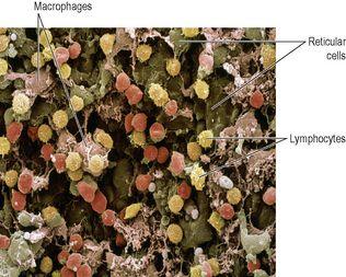
Figure 6.6
shows a scanning electron micrograph of lymph node tissue, with reticular cells, white blood cells and macrophages.
Figure 6.6
Colour scanning electron micrograph of lymph node tissue
. Cell population includes reticular cells (brown), macrophages (pink) and lymphocytes (yellow).
Spleen
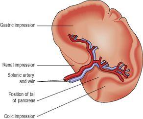
The spleen (
Fig. 6.7
) contains reticular and lymphatic tissue and is the largest lymph organ.
Figure 6.7
The spleen.
The spleen lies in the left hypochondriac region of the abdominal cavity between the fundus of the stomach and the diaphragm. It is purplish in colour and varies in size in different individuals, but is usually about 12 cm long, 7 cm wide and 2.5 cm thick. It weighs about 200 g.
Organs associated with the spleen
Superiorly and posteriorly
– diaphragm
Inferiorly
– left colic flexure of the large intestine
Anteriorly
– fundus of the stomach
Medially
– pancreas and the left kidney
Laterally
– separated from the 9th, 10th and 11th ribs and the intercostal muscles by the diaphragm
Structure (
Fig. 6.8
)
The spleen is slightly oval in shape with the hilum on the lower medial border. The anterior surface is covered with peritoneum. It is enclosed in a fibroelastic capsule that dips into the organ, forming trabeculae. The cellular material, consisting of lymphocytes and macrophages, is called
splenic pulp
, and lies between the trabeculae.
Red pulp
is the part suffused with blood and
white pulp
consists of areas of lymphatic tissue where there are sleeves of lymphocytes and macrophages around blood vessels.
Figure 6.8
A section through the spleen.
The structures entering and leaving the spleen at the hilum are:
•
splenic artery, a branch of the coeliac artery
•
splenic vein, a branch of the portal vein
•
lymph vessels (efferent only)
•
nerves.
Blood passing through the spleen flows in sinusoids (
p. 75
), which have distinct pores between the endothelial cells, allowing it to come into close association with splenic pulp.
Functions
Phagocytosis
As described previously (
p. 60
), old and abnormal erythrocytes are mainly destroyed in the spleen, and the breakdown products, bilirubin and iron, are transported to the liver via the splenic and portal veins. Other cellular material, e.g. leukocytes, platelets and bacteria, is phagocytosed in the spleen. Unlike lymph nodes, the spleen has no afferent lymphatics entering it, so it is not exposed to diseases spread by lymph.
Storage of blood
The spleen contains up to 350 ml of blood, and in response to sympathetic stimulation can rapidly return most of this volume to the circulation, e.g. in haemorrhage.
Immune response
The spleen contains T- and B-lymphocytes, which are activated by the presence of antigens, e.g. in infection. Lymphocyte proliferation during serious infection can cause enlargement of the spleen (
splenomegaly
).
Erythropoiesis
The spleen and liver are important sites of fetal blood cell production, and the spleen can also fulfil this function in adults in times of great need.
Thymus gland


