Ross & Wilson Anatomy and Physiology in Health and Illness (28 page)
Read Ross & Wilson Anatomy and Physiology in Health and Illness Online
Authors: Anne Waugh,Allison Grant
Tags: #Medical, #Nursing, #General, #Anatomy

discuss the functions and formation of the different types of white blood cell
outline the role of platelets in blood clotting.
There are three types of blood cell (see
Fig. 1.6, p. 8
).
•
erythrocytes (red cells)
•
platelets (thrombocytes)
•
leukocytes (white cells).
Blood cells are synthesised mainly in red bone marrow. Some lymphocytes, additionally, are produced in lymphoid tissue. In the bone marrow, all blood cells originate from
pluripotent
(i.e. capable of developing into one of a number of cell types)
stem cells
and go through several developmental stages before entering the blood. Different types of blood cell follow separate lines of development. The process of blood cell formation is called
haemopoiesis
(
Fig. 4.2
).
Figure 4.2
Haemopoiesis:
stages in the development of blood cells.
For the first few years of life, red marrow occupies the entire bone capacity and, over the next 20 years, is gradually replaced by fatty yellow marrow that has no haemopoietic function. In adults, haemopoiesis in the skeleton is confined to flat bones, irregular bones and the ends (
epiphyses
) of long bones, the main sites being the sternum, ribs, pelvis and skull.
Erythrocytes (red blood cells)
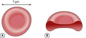
Red blood cells are biconcave discs; they have no nucleus, and their diameter is about 7 micrometres (
Fig. 4.3
). Their main function is in gas transport, mainly of oxygen, but they also carry some carbon dioxide. Their characteristic shape is suited to their purpose; the biconcavity increases their surface area for gas exchange, and the thinness of the central portion allows fast entry and exit of gases. The cells are flexible so they can squeeze through narrow capillaries, and contain no intracellular organelles, leaving more room for haemoglobin, the large pigmented protein responsible for gas transport.
Figure 4.3
A
and
B.
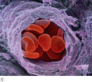
The red blood cell.
C.
Coloured scanning electron micrograph of a group of red blood cells travelling along an arteriole.
Measurements of red cell numbers, volume and haemoglobin content are routine and useful assessments made in clinical practice (
Table 4.1
). The symbols in brackets are the abbreviations commonly used in laboratory reports.
Table 4.1
Erythrocytes – normal values
| Measure | Normal values |
|---|---|
| Erythrocyte count – number of erythrocytes per litre, or cubic millilitre, (mm 3 ) of blood | Male: 4.5 × 10 12 /l to 6.5 × 10 12 /l (4.5–6.5 million/mm 3 ) |
| Female: 3.8 × 10 12 /l to 5.8 ×10 12 /l (3.8–5.8 million/mm 3 ) | |
| Packed cell volume (PCV, haematocrit) – the volume of red cells in 1 l or mm 3 of blood | 0.40–0.55 l/l |
| Mean cell volume (MCV) – the volume of an average cell, measured in femtolitres (1 fl = 10 −15 litre) | 80–96 fl |
| Haemoglobin – the weight of haemoglobin in whole blood, measured in grams/100 ml blood | Male: 13–18 g/100 ml |
| Female: 11.5–16.5 g/100 ml | |
| Mean cell haemoglobin (MCH) – the average amount of haemoglobin per cell, measured in picograms (1 pg = 10 −12 gram) | 27–32 pg/cell |
| Mean cell haemoglobin concentration (MCHC) – the weight of haemoglobin in 100 ml of red cells | 30–35 g/100 ml of red cells |
Life span and function of erythrocytes
Erythrocytes are produced in red bone marrow, which is present in the ends of long bones and in flat and irregular bones. They pass through several stages of development before entering the blood. Their life span in the circulation is about 120 days.
The process of development of red blood cells from stem cells takes about 7 days and is called
erythropoiesis
(
Fig. 4.2
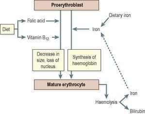
). The immature cells are released into the bloodstream as reticulocytes, and then mature into erythrocytes over a day or two within the circulation. During this time, they lose their nucleus and therefore become incapable of division (
Fig. 4.4
).
Figure 4.4
Maturation of the erythrocyte.
Both vitamin B
12
and folic acid are required for red blood cell synthesis. They are absorbed in the intestines, although vitamin B
12
must be bound to intrinsic factor (
p. 292
) to allow absorption to take place. Both vitamins are present in dairy products, meat and green vegetables. The liver usually contains substantial stores of vitamin B
12
, several years’ worth, but signs of folic acid deficiency appear within a few months.
Haemoglobin
Haemoglobin is a large, complex protein containing a globular protein (globin) and a pigmented iron-containing complex called haem. Each haemoglobin molecule contains four globin chains and four haem units, each with one atom of iron (
Fig. 4.5
). As each atom of iron can combine with an oxygen molecule, this means that a single haemoglobin molecule can carry up to four molecules of oxygen. An average red blood cell carries about 280 million haemoglobin molecules, giving each cell a theoretical oxygen-carrying capacity of over a billion oxygen molecules!


