Ross & Wilson Anatomy and Physiology in Health and Illness (23 page)
Read Ross & Wilson Anatomy and Physiology in Health and Illness Online
Authors: Anne Waugh,Allison Grant
Tags: #Medical, #Nursing, #General, #Anatomy

This part of the chapter provides an overview of anatomical terms and the names and positions of bones. A more detailed account of the bones, muscles and joints is given in
Chapter 16
.
Anatomical terms
The anatomical position
This is the position assumed in all anatomical descriptions to ensure accuracy and consistency. The body is in the upright position with the head facing forward, the arms at the sides with the palms of the hands facing forward and the feet together.
Median plane
When the body, in the anatomical position, is divided
longitudinally
through the midline into right and left halves it has been divided in the median plane.
Directional terms
These paired terms are used to describe the location of body parts in relation to others, and are explained in
Table 3.1
.
Table 3.1
Paired directional terms used in anatomy
| Directional term | Meaning |
|---|---|
| Medial | Structure is nearer to the midline. The heart is medial to the humerus |
| Lateral | Structure is further from the midline or at the side of the body. The humerus is lateral to the heart |
| Proximal | Nearer to a point of attachment of a limb, or origin of a body part. The femur is proximal to the fibula |
| Distal | Further from a point of attachment of a limb, or origin of a body part. The fibula is distal to the femur |
| Anterior or ventral | Part of the body being described is nearer the front of the body. The sternum is anterior to the vertebrae |
| Posterior or dorsal | Part of the body being described is nearer the back of the body. The vertebrae are posterior to the sternum |
| Superior | Structure nearer the head. The skull is superior to the scapulae |
| Inferior | Structure further from the head. The scapulae are inferior to the skull |
Regional terms
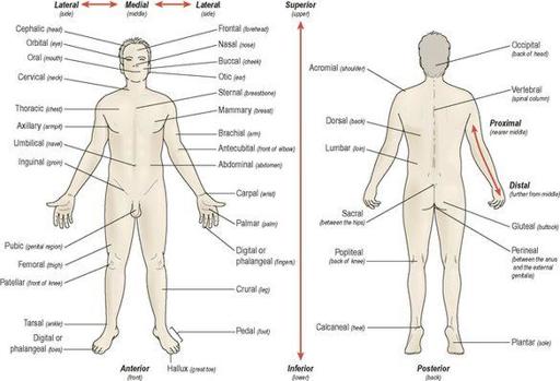
These are used to describe parts of the body (
Fig. 3.26
).
Figure 3.26
Regional and directional terms.
The skeleton
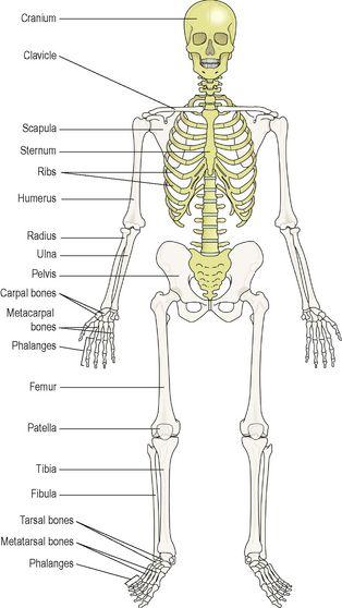
The skeleton (
Fig. 3.27
) is the bony framework of the body. It forms the cavities and fossae (depressions or hollows) that protect some structures, forms the joints and gives attachment to muscles. A detailed description of the bones is given in
Chapter 16
.
Table 16.1
,
page 385
lists the terminology related to the skeleton.
Figure 3.27
Anterior view of the skeleton:
axial skeleton – gold, appendicular skeleton – brown.
The skeleton is described in two parts:
axial
and
appendicular
(the appendages attached to the axial skeleton).
Axial skeleton
The axial skeleton (axis of the body) consists of the skull, vertebral column, sternum (or breast bone) and the ribs.
Skull
The skull is described in two parts, the
cranium
, which contains the brain, and the
face
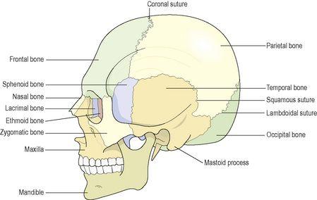
. It consists of a number of bones, which develop separately but fuse together as they mature. The only movable bone is the mandible or lower jaw. The names and positions of the individual bones of the skull can be seen in
Figure 3.28
.
Figure 3.28
The skull: bones of the cranium and face.
Functions of the skull
The various parts of the skull have specific and different functions (see
p. 391
) and are, in summary:
•
protection of delicate structures including the brain, eyes and inner ears
•
maintaining patency of the nasal passages enabling breathing
•
eating – the teeth are embedded in the mandible and maxilla; and movement of the mandible, the only movable skull bone, allows chewing.
Vertebral column
This consists of 24 movable bones (vertebrae) plus the sacrum and coccyx. The bodies of the bones are separated from each other by
intervertebral discs
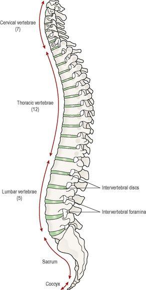
, consisting of cartilage. The vertebral column is described in five parts and the bones of each part are numbered from above downwards (
Fig. 3.29
):
•
7 cervical
•
12 thoracic
•
5 lumbar
•
1 sacrum (5 fused bones)
•
1 coccyx (4 fused bones).
Figure 3.29
The vertebral column – lateral view.
The first cervical vertebra, called the
atlas
, forms a joint (
articulates
) with the skull. Thereafter each vertebra forms a joint with the vertebrae immediately above and below. More movement is possible in the cervical and lumbar regions than in the thoracic region.
The
sacrum
consists of five vertebrae fused into one bone that articulates with the fifth lumbar vertebra above, the coccyx below and an innominate (pelvic or hip) bone at each side.
The
coccyx
consists of the four terminal vertebrae fused into a small triangular bone that articulates with the sacrum above.
Functions of the vertebral column
The vertebral column has several important functions:
•
It protects the spinal cord. In each vertebra is a hole, the
vertebral foramen
, and collectively the foramina form a canal in which the spinal cord lies.
•
Adjacent vertebrae form openings (intervertebral foramina), which protect the spinal nerves as they pass from the spinal cord (see
Fig. 16.26, p. 394
).
•
In the thoracic region the ribs articulate with the vertebrae forming joints allowing movement of the ribcage during respiration.
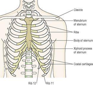
Thoracic cage
The thoracic cage is formed by:
•
12 thoracic vertebrae
•
12 pairs of ribs
•
1 sternum or breast bone.
The arrangement of the bones is shown in
Figure 3.30
.
Figure 3.30
The structures forming the walls of the thoracic cage.

