Ross & Wilson Anatomy and Physiology in Health and Illness (22 page)
Read Ross & Wilson Anatomy and Physiology in Health and Illness Online
Authors: Anne Waugh,Allison Grant
Tags: #Medical, #Nursing, #General, #Anatomy

•
tissues in which cell replication is a continuous process regenerate quickly – these include epithelial cells of, for example, the skin, mucous membrane, secretory glands, uterine lining and lymphoid tissue
•
other tissues retain the ability to replicate, but do so infrequently; these include the liver, kidney, fibroblasts and smooth muscle cells. These tissues take longer to regenerate
•
some tissues are normally unable to replicate including nerve cells (neurones) and skeletal and cardiac muscle cells. Extensively damaged tissue is usually replaced by fibrous tissue, meaning that the functions carried out by the original tissue are lost.
Membranes
Epithelial membranes
These membranes are sheets of epithelial tissue and supporting connective tissue that cover or line many internal structures or cavities. The main ones are mucous membrane, serous membrane and the skin (cutaneous membrane, see
Ch. 14
).
Mucous membrane
This is the moist lining of the alimentary, respiratory and genitourinary tracts which is sometimes referred to as the
mucosa
. The membrane surface consists of epithelial cells, some of which produce a secretion called
mucus
, a slimy tenacious fluid. As it accumulates the cells become distended and finally burst, discharging the mucus onto the free surface. As the cells fill up with mucus they have the appearance of a goblet or flask and are known as
goblet cells
(see
Fig. 12.5, p. 282
). Organs lined by mucous membrane have a moist slippery surface. Mucus protects the lining membrane from drying, and mechanical and chemical injury. In the respiratory tract it traps inhaled foreign particles, preventing them from entering the alveoli of the lungs.
Serous membrane
Serous membranes, or
serosa
, secrete serous watery fluid. They consist of a double layer of loose areolar connective tissue lined by simple squamous epithelium. The
parietal
layer lines a cavity and the
visceral
layer surrounds organs (the viscera) within the cavity. The two layers are separated by
serous fluid
secreted by the epithelium. There are three sites where serous membranes are found:
•
the
pleura
lining the thoracic cavity and surrounding the lungs (
p. 243
)
•
the
pericardium
lining the pericardial cavity and surrounding the heart (
p. 79
)
•
the
peritoneum
lining the abdominal cavity and surrounding abdominal organs (
p. 280
).
The serous fluid between the visceral and parietal layers enables an organ to glide freely within the cavity without being damaged by friction between it and adjacent organs. For example, the heart changes its shape and size during each beat and friction damage is prevented by the arrangement of pericardium and its serous fluid.
Synovial membrane
This membrane lines the cavities of moveable joints and surrounds tendons that could be injured by rubbing against bones, e.g. over the wrist joint. It is not an epithelial membrane, but instead consists of areolar connective tissue and elastic fibres.
Synovial membrane secretes clear, sticky, oily
synovial fluid
, which lubricates and nourishes the joints (see
Ch. 16
).
Glands
Glands
are groups of epithelial cells that produce specialised secretions. Glands that discharge their secretion onto the epithelial surface of hollow organs, either directly or through a
duct
, are called
exocrine glands
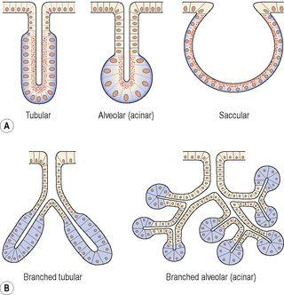
. Exocrine glands vary considerably in size, shape and complexity as shown in
Figure 3.24
. Secretions of exocrine glands include mucus, saliva, digestive juices and earwax.
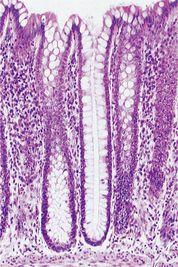
Figure 3.25
shows simple tubular glands of the large intestine. Other glands discharge their secretions into blood and lymph. These are called
endocrine glands
(ductless glands) and their secretions are
hormones
(see
Ch. 9
).
Figure 3.24
Exocrine glands: A.
Simple glands.
B.
Compound (branching) glands.
Figure 3.25
Simple tubular glands in the large intestine. A stained photograph (magnified × 50).
Organisation of the body
Learning outcomes
After studying this section you should be able to:
define common anatomical terms
identify the principal bones of the axial skeleton and the appendicular skeleton
state the boundaries of the four body cavities
list the contents of the body cavities.


