Ross & Wilson Anatomy and Physiology in Health and Illness (17 page)
Read Ross & Wilson Anatomy and Physiology in Health and Illness Online
Authors: Anne Waugh,Allison Grant
Tags: #Medical, #Nursing, #General, #Anatomy

Figure 3.2
The plasma membrane. A.
Diagram showing structure.
B.
Coloured atomic force micrograph of the surface showing plasma proteins.
The phospholipid molecules have a head, which is electrically charged and
hydrophilic
(meaning ‘water loving’), and a tail which has no charge and is
hydrophobic
(meaning ‘water hating’,
Fig. 3.2A
). The phospholipid bilayer is arranged like a sandwich with the hydrophilic heads aligned on the outer surfaces of the membrane and the hydrophobic tails forming a central water-repelling layer. These differences influence the transfer of substances across the membrane.
The membrane proteins perform several functions:
•
branched carbohydrate molecules attached to the outside of some membrane protein molecules give the cell its immunological identity
•
they can act as specific receptors (recognition sites) for hormones and other chemical messengers
•
some are enzymes (
p. 24
)
•
some are involved in transport across the membrane.
Organelles
Nucleus
Every cell in the body has a nucleus, with the exception of mature erythrocytes (red blood cells). Skeletal muscle and some other cells contain several nuclei. The nucleus is the largest organelle and is contained within the nuclear envelope, a membrane similar to the plasma membrane but with tiny pores through which some substances can pass between it and the
cytoplasm
, i.e. the cell contents excluding the nucleus.
The nucleus contains the body’s genetic material, which directs all the metabolic activities of the cell. This consists of 46
chromosomes
, which are made from deoxyribonucleic acid (DNA,
p. 428
). Except during cell division, the chromosomes resemble a fine network of threads called
chromatin
.
Within the nucleus is a roughly spherical structure called the
nucleolus,
which is involved in manufacture (synthesis) and assembly of the components of ribosomes.
Mitochondria
Mitochondria are membranous, sausage-shaped structures in the cytoplasm, sometimes described as the ‘power house’ of the cell (
Fig. 3.3
). They are involved in aerobic respiration, the processes by which chemical energy is made available in the cell. This is in the form of ATP, which releases energy when the cell breaks it down (see
Fig. 2.10, p. 24
). Synthesis of ATP is most efficient in the final stages of aerobic respiration, a process requiring oxygen (
p. 308
). The most active cell types have the greatest number of mitochondria, e.g. liver, muscle and spermatozoa.
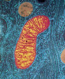
Figure 3.3
Mitochondrion and rough endoplasmic reticulum.
False colour transmission electron micrograph showing mitochondrion (orange) and rough endoplasmic reticulum (turquoise) studded with ribosomes (dots).
Ribosomes
These are tiny granules composed of RNA and protein. They synthesise proteins from amino acids, using RNA as the template (see
Fig. 17.5, p. 430
). When present in free units or in small clusters in the cytoplasm, the ribosomes make proteins for use within the cell. These include the enzymes required for metabolism. Metabolic pathways consist of a series of steps, each driven by a specific enzyme. Ribosomes are also found on the outer surface of the nuclear envelope and rough endoplasmic reticulum (see
Fig. 3.3
and below) where they manufacture proteins for export from the cell.
Endoplasmic reticulum (ER)
Endoplasmic reticulum is an extensive series of interconnecting membranous canals in the cytoplasm (
Fig. 3.3
). There are two types: smooth and rough. Smooth ER synthesises lipids and steroid hormones, and is also associated with the detoxification of some drugs. Some of the lipids are used to replace and repair the plasma membrane and membranes of organelles. Rough ER is studded with ribosomes. These are the site of synthesis of proteins, some of which are ‘exported’ from cells, i.e. enzymes and hormones that leave the parent cell by exocytosis (
p. 33
) to be used by cells elsewhere.
Golgi apparatus
The Golgi apparatus consists of stacks of closely folded flattened membranous sacs (
Fig. 3.4
). It is present in all cells but is larger in those that synthesise and export proteins. The proteins move from the endoplasmic reticulum to the Golgi apparatus where they are ‘packaged’ into membrane-bound vesicles called
secretory granules
. The vesicles are stored and, when needed, they move to the plasma membrane and fuse with it. The contents then leave the cell by exocytosis (
p. 33
).
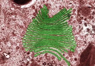
Figure 3.4
Coloured transmission electron micrograph showing the Golgi apparatus (green).
Lysosomes
Lysosomes are one type of secretory vesicle with membranous walls, which are formed by the Golgi apparatus. They contain a variety of enzymes involved in breaking down fragments of organelles and large molecules (e.g. RNA, DNA, carbohydrates, proteins) inside the cell into smaller particles that are either recycled, or extruded from the cell as waste material.
Lysosomes in white blood cells contain enzymes that digest foreign material such as microbes.
Cytoskeleton
This consists of an extensive network of tiny protein fibres (
Fig. 3.5
).
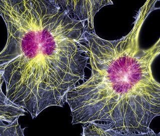
Figure 3.5
Fibroblasts.
Fluorescent light micrograph showing their nuclei (purple) and cytoskeletons (yellow and blue).
Microfilaments
These are the smallest fibres. They provide structural support, maintain the characteristic shape of the cell and permit contraction, e.g. in muscle cells.
Microtubules
These are larger contractile protein fibres that are involved in movement of:
•
organelles within the cell
•
chromosomes during cell division
•
cell extensions (see below).
Centrosome
This directs organisation of microtubules within the cell. It consists of a pair of
centrioles
(small clusters of microtubules) and plays an important role during cell division.
Cell extensions
These project from the plasma membrane in some types of cell and their main components are microtubules, which allow movement. They include:
•
microvilli – tiny projections that contain microfilaments. They cover the surface of certain types of cell, e.g. absorptive cells that line the small intestine (see
Fig. 3.6
). By greatly increasing the surface area, microvilli make the structure of these cells ideal for their function – maximising absorption of nutrients from the small intestine.
•
cilia – microscopic hair-like projections containing microtubules that lie along the free borders of some cells (see
Fig. 10.12, p. 241
). They beat in unison, moving substances along the surface, e.g. mucus upwards in the respiratory tract.
•
flagella – single, long whip-like projections, containing microtubules, which form the ‘tails’ of spermatozoa (see
Fig. 1.17, p. 14
) that enable their movement along the female reproductive tract.
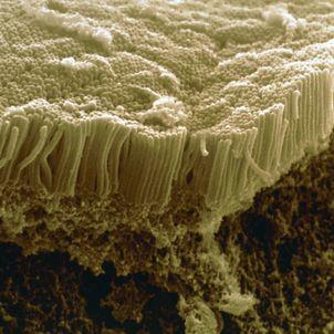
Figure 3.6
Coloured scanning electron micrograph of microvilli in small intestine.
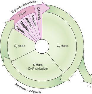
The cell cycle
Many damaged, dead, and worn out cells can be replaced by growth and division of other similar cells. Most body cells have 46 chromosomes and divide by
mitosis,
a process that results in two new genetically identical daughter cells. The only exception to this is the formation of
gametes
(sex cells), i.e. ova and spermatozoa, which takes place by
meiosis
(
p. 432
).
The period between two cell divisions is known as the
cell cycle,
which has two phases that can be seen on light microscopy: mitosis (M phase) and
interphase
(
Fig. 3.7
).