Ross & Wilson Anatomy and Physiology in Health and Illness (16 page)
Read Ross & Wilson Anatomy and Physiology in Health and Illness Online
Authors: Anne Waugh,Allison Grant
Tags: #Medical, #Nursing, #General, #Anatomy

Axial skeleton
43
Appendicular skeleton
45
Cavities of the body
45
Cranial cavity
45
Thoracic cavity
46
Abdominal cavity
46
Pelvic cavity
47
Disorders of cells and tissues
49
Neoplasms or tumours
49
Causes of neoplasms
49
Growth of tumours
50
Effects of tumours
51
Causes of death in malignant disease
51
ANIMATIONS
3.1
Cellular functions compared to organ systems
28
3.2
Mitosis
31
3.3
Selective permeability
31
3.4
Passive transport
32
3.5
Active transport
32
3.6
Mucous membrane
40
3.7
Serous membrane
40
3.8
Synovial membrane
40
3.9
Vertebral column
43
3.10
Anatomy and physiology of the abdomen
46
Cells
are the smallest functional units of the body. They are grouped together to form
tissues
, each of which has a specialised function, e.g. blood, muscle, bone. Different tissues are grouped together to form
organs
, e.g. heart, stomach, brain. Organs are grouped together to form
systems
, each of which performs a particular function that maintains homeostasis and contributes to the health of the individual (see
Fig. 1.2, p. 5
). For example, the digestive system is responsible for taking in, digesting and absorbing food and involves a number of organs, including the stomach and intestines. The structure and functions of cells and types of tissue are explored in this chapter.
The terminology used to describe the anatomical relationships of body parts, the skeleton and the cavities within the body are then described.
The final section considers features of benign and malignant tumours, their causes and how they grow and may spread.
The cell: structure and functions

Learning outcomes
After studying this section you should be able to:
describe the structure of the plasma membrane
The human body develops from a single cell called the
zygote
, which results from the fusion of the ovum (female egg cell) and the spermatozoon (male sex cell). Cell division follows and, as the fetus grows, cells with different structural and functional specialisations develop, all with the same genetic make-up as the zygote. Individual cells are too small to be seen with the naked eye. However, they can be seen when thin slices of tissue are stained in the laboratory and magnified by a microscope.
A cell consists of a
plasma membrane
inside which are a number of
organelles
suspended in a watery fluid called
cytoplasm
(
Fig. 3.1
). Organelles, literally ‘small organs’, have individual and highly specialised functions, and are often enclosed in their own membrane within the cytoplasm. They include: the
nucleus
,
mitochondria
,
ribosomes
,
endoplasmic reticulum
,
Golgi apparatus
,
lysosomes
and the
cytoskeleton
.

Figure 3.1
The simple cell.
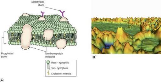
Plasma membrane
The plasma membrane (
Fig. 3.2
) consists of two layers of
phospholipids
(fatty substances, see
p. 23
) with protein and sugar molecules embedded in them. In addition to phospholipids, the lipid
cholesterol
is also present in the plasma membrane. Those proteins that extend all the way through the membrane may provide channels that allow the passage of, for example, electrolytes and non-lipid-soluble substances. Protein molecules on the surface of the plasma membrane are shown in
Figure 3.2B
.