Ross & Wilson Anatomy and Physiology in Health and Illness (19 page)
Read Ross & Wilson Anatomy and Physiology in Health and Illness Online
Authors: Anne Waugh,Allison Grant
Tags: #Medical, #Nursing, #General, #Anatomy

Figure 3.11
Bulk transport across plasma membranes: A–E.
Phagocytosis.
F.
Exocytosis.
Export of waste material by the reverse process through the plasma membrane is called
exocytosis
. Secretory granules formed by the Golgi apparatus usually leave the cell in this way, as do any indigestible residues of phagocytosis.
Tissues
Learning outcomes
After studying this section you should be able to:
describe the structure and functions of epithelial, connective and muscle tissue
outline the structure and functions of membranes
compare and contrast the structure and functions of exocrine and endocrine glands.
The tissues of the body consist of large numbers of cells and they are classified according to the size, shape and functions of these cells. There are four main types of tissue that each have subdivisions. They are:
•
epithelial tissue or epithelium
•
connective tissue
•
muscle tissue
•
nervous tissue.
Epithelial tissue (
Fig. 3.12
)
This group of tissues is found covering the body and lining cavities, hollow organs and tubes. It is also found in glands. The structure of epithelium is closely related to its functions, which include:
•
protection of underlying structures from, for example, dehydration, chemical and mechanical damage
•
secretion
•
absorption.
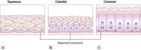
Figure 3.12
Simple epithelium. A.
Squamous.
B.
Cuboidal.
C.
Columnar.
The cells are very closely packed and the intercellular substance, called the
matrix
, is minimal. The cells usually lie on a
basement membrane
, which is an inert connective tissue made by the epithelial cells themselves.
Epithelial tissue may be:
•
simple
: a single layer of cells
•
stratified
: several layers of cells.
Simple epithelium
Simple epithelium consists of a single layer of identical cells and is divided into three main types. It is usually found on absorptive or secretory surfaces, where the single layer enhances these processes, and not usually on surfaces subject to stress. The types are named according to the shape of the cells, which differs according to their functions. The more active the tissue, the taller the cells.
Squamous (pavement) epithelium
This is composed of a single layer of flattened cells (
Fig. 3.12A
). The cells fit closely together like flat stones, forming a thin and very smooth membrane across which diffusion easily occurs. It forms the lining of the following structures:
•
heart – where it is known as endocardium

•
alveoli of the lungs
•
lining the collecting ducts of nephrons in the kidneys (see
Fig. 13.9, p. 333
).
Cuboidal epithelium
This consists of cube-shaped cells fitting closely together lying on a basement membrane (
Fig. 3.12B
). It forms the kidney tubules and is found in some glands. Cuboidal epithelium is actively involved in secretion, absorption and excretion.
Columnar epithelium
This is formed by a single layer of cells, rectangular in shape, on a basement membrane (
Fig. 3.12C
). It lines many organs and often has adaptations that make it well suited to a specific function. The lining of the stomach is formed from simple columnar epithelium without surface structures. The surface of the columnar epithelium lining the small intestine is covered with microvilli (
Fig. 3.6
). Microvilli provide a very large surface area for absorption of nutrients from the small intestine. In the trachea, columnar epithelium is ciliated (see
Fig. 10.12, p. 241
) and also contains goblet cells that secrete mucus (see
Fig. 12.5, p. 282
). This means that inhaled particles that stick to the mucus layer are moved towards the throat by cilia (
p. 241
) in the respiratory tract. In the uterine tubes, ova are propelled along by ciliary action towards the uterus.
Stratified epithelia
Stratified epithelia consist of several layers of cells of various shapes. Continual cell division in the lower (basal) layers pushes cells above nearer and nearer to the surface, where they are shed. Basement membranes are usually absent. The main function of stratified epithelium is to protect underlying structures from mechanical wear and tear. There are two main types: stratified squamous and transitional.
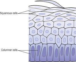
Stratified squamous epithelium (
Fig. 3.13
)
This is composed of a number of layers of cells. In the deepest layers the cells are mainly columnar and, as they grow towards the surface, they become flattened and are then shed.
Figure 3.13
Stratified epithelium.
Keratinised stratified epithelium
This is found on dry surfaces subjected to wear and tear, i.e. skin, hair and nails. The surface layer consists of dead epithelial cells that have lost their nuclei and contain the protein keratin. This forms a tough, relatively waterproof protective layer that prevents drying of the live cells underneath. The surface layer of skin is rubbed off and is replaced from below (see
Ch. 14
).
Non-keratinised stratified epithelium
This protects moist surfaces subjected to wear and tear, and prevents them from drying out, e.g. the conjunctiva of the eyes, the lining of the mouth, the pharynx, the oesophagus and the vagina (
Fig. 3.14
).


