Ross & Wilson Anatomy and Physiology in Health and Illness (21 page)
Read Ross & Wilson Anatomy and Physiology in Health and Illness Online
Authors: Anne Waugh,Allison Grant
Tags: #Medical, #Nursing, #General, #Anatomy

•
as an outer protective covering for bone, called
periosteum
•
as an outer protective covering of some organs, e.g. the kidneys, lymph nodes and the brain
•
forming muscle sheaths, called
muscle fascia
(see
Fig. 16.52
,
p. 408
), which extend beyond the muscle to become the
tendon
that attaches the muscle to bone.
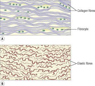
Elastic tissue (
Fig. 3.19B
)
Elastic tissue is capable of considerable extension and recoil. There are few cells and the matrix consists mainly of masses of elastic fibres secreted by fibroblasts. It is found in organs where stretching or alteration of shape is required, e.g. in large blood vessel walls, the trachea and bronchi, and the lungs.
Figure 3.19
Dense connective tissue. A.
Fibrous tissue.
B.
Elastic tissue.
Blood
This is a fluid connective tissue that is described in detail in
Chapter 4
.
Cartilage
Cartilage is firmer than other connective tissues; the cells are called
chondrocytes
and are less numerous. They are embedded in matrix reinforced by collagen and elastic fibres. There are three types: hyaline cartilage, fibrocartilage and elastic fibrocartilage.
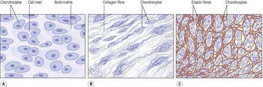
Hyaline cartilage (
Fig. 3.20A
)
Hyaline cartilage is a smooth bluish-white tissue. The chondrocytes are in small groups within cell nests and the matrix is solid and smooth. Hyaline cartilage provides flexibility, support and smooth surfaces for movement at joints. It is found:
•
on the ends of long bones that form joints
•
forming the costal cartilages, which attach the ribs to the sternum
•
forming part of the larynx, trachea and bronchi.
Figure 3.20
Cartilage. A.
Hyaline cartilage.
B.
Fibrocartilage.
C.
Elastic fibrocartilage.
Fibrocartilage (
Fig. 3.20B
)
This consists of dense masses of white collagen fibres in a matrix similar to that of hyaline cartilage with the cells widely dispersed. It is a tough, slightly flexible, supporting tissue found:
•
as pads between the bodies of the vertebrae, the
intervertebral discs
•
between the articulating surfaces of the bones of the knee joint, called
semilunar cartilages
•
on the rim of the bony sockets of the hip and shoulder joints, deepening the cavities without restricting movement
•
as
ligaments
joining bones.
Elastic fibrocartilage (
Fig. 3.20C
)
This flexible tissue consists of yellow elastic fibres lying in a solid matrix. The chondrocytes lie between the fibres. It provides support and maintains shape of, e.g. the pinna or lobe of the ear, the epiglottis and part of the tunica media of blood vessel walls.
Bone
Bone cells (osteocytes) are surrounded by a matrix of collagen fibres strengthened by inorganic salts, especially calcium and phosphate. This provides bones with their characteristic strength and rigidity. Bone also has considerable capacity for growth in the first two decades of life, and for regeneration throughout life. Two types of bone can be identified by the naked eye:
•
compact bone
– solid or dense appearance
•
spongy
or
cancellous bone
– ‘spongy’ or fine honeycomb appearance.
These are described in detail in
Chapter 16
.
Muscle tissue
Muscle tissue is able to contract and relax, providing movement within the body and of the body itself. Muscle contraction requires an adequate blood supply to provide sufficient oxygen, calcium and nutrients and to remove waste products. There are three types of specialised contractile cells, also known as
fibres
: skeletal muscle, smooth muscle and cardiac muscle.
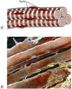
Skeletal muscle tissue (
Fig. 3.21
)
This type is described as skeletal because it forms those muscles that move the bones [of the skeleton],
striated
because striations (stripes) can be seen on microscopic examination and
voluntary
as it is under conscious control. In reality, movements can be finely coordinated, e.g. writing, but may also be controlled subconsciously. For example, maintaining an upright posture does not normally require thought unless a new locomotor skill is being learned, e.g. skating or cycling, and the diaphragm maintains breathing while asleep.
Figure 3.21
Skeletal muscle fibres. A.
Diagram.
B.
Coloured scanning electron micrograph of skeletal muscle fibres and connective tissue fibres (bottom right).
Fibres are cylindrical, contain several nuclei and can be up to 35 cm long. Skeletal muscle contraction is stimulated by motor nerve impulses originating in the brain or spinal cord and ending at the neuromuscular junction (see
p. 411
). The properties and functions of skeletal muscle are explained in detail in
Chapter 16
.
Smooth muscle tissue (
Fig. 3.22
)
Smooth muscle may also be described as
non-striated, visceral
or
involuntary
. It does not have striations and is not under conscious control. Smooth muscle has the intrinsic ability to contract and relax. Additionally, autonomic nerve impulses, some hormones and local metabolites stimulate contraction. A degree of muscle tone is always present, meaning that smooth muscle is completely relaxed for only short periods. Contraction of smooth muscle is slower and more sustained than skeletal muscle. It is found in the walls of hollow organs:
•
regulating the diameter of blood vessels and parts of the respiratory tract
•
propelling contents of the ureters, ducts of glands and alimentary tract
•
expelling contents of the urinary bladder and uterus.
Figure 3.22.
Smooth muscle. A.
Diagram.
B.
Fluorescent light micrograph showing actin, a contractile muscle protein (green), nuclei (blue) and capillaries (red).
When examined under a microscope, the cells are seen to be spindle shaped with only one central nucleus. Bundles of fibres form sheets of muscle, such as those found in the walls of the above structures.
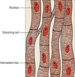
Cardiac muscle tissue (
Fig. 3.23
)
This type of muscle tissue is found only in the heart wall. It is not under conscious control but, when viewed under a microscope, cross-stripes (striations) characteristic of skeletal muscle can be seen. Each fibre (cell) has a nucleus and one or more branches. The ends of the cells and their branches are in very close contact with the ends and branches of adjacent cells. Microscopically these ‘joints’, or
intercalated discs
, can be seen as lines that are thicker and darker than the ordinary cross-stripes. This arrangement gives cardiac muscle the appearance of a sheet of muscle rather than a very large number of individual fibres. The end-to-end continuity of cardiac muscle cells has significance in relation to the way the heart contracts. A wave of contraction spreads from cell to cell across the intercalated discs, which means that cells do not need to be stimulated individually.
Figure 3.23
Cardiac muscle fibres.
The heart has an intrinsic pacemaker system, which means that it beats in a coordinated manner without external nerve stimulation, although the rate at which it beats is influenced by autonomic nerve impulses, some hormones, local metabolites and other substances (see
Ch. 5
).
Nervous tissue
Two types of tissue are found in the nervous system:
•
excitable cells – these are called
neurones
and they initiate, receive, conduct and transmit information
•
non-excitable cells – also known as
glial cells
, these support the neurones.
These are described in detail in
Chapter 7
.
Tissue regeneration
The extent to which regeneration is possible depends on the normal rate of turnover of particular types of cell. Those with a rapid turnover regenerate most effectively. There are three general categories:


