Ross & Wilson Anatomy and Physiology in Health and Illness (61 page)
Read Ross & Wilson Anatomy and Physiology in Health and Illness Online
Authors: Anne Waugh,Allison Grant
Tags: #Medical, #Nursing, #General, #Anatomy

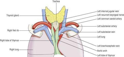
The thymus gland lies in the upper part of the mediastinum behind the sternum and extends upwards into the root of the neck (
Fig. 6.9
). It weighs about 10 to 15 g at birth and grows until puberty, when it begins to atrophy. Its maximum weight, at puberty, is between 30 and 40 g and by middle age it has returned to approximately its weight at birth.
Figure 6.9
The thymus gland in the adult, and related structures.
Organs associated with the thymus
Anteriorly
– sternum and upper four costal cartilages
Posteriorly
– aortic arch and its branches, brachiocephalic veins, trachea
Laterally
– lungs
Superiorly
– structures in the root of the neck
Inferiorly
– heart
Structure
The thymus consists of two lobes joined by areolar tissue. The lobes are enclosed by a fibrous capsule which dips into their substance, dividing them into lobules that consist of an irregular branching framework of epithelial cells and lymphocytes.
Function
Lymphocytes originate from stem cells in red bone marrow (
p. 58
). Those that enter the thymus develop into activated T-lymphocytes (
p. 370
).
Thymic processing produces mature T-lymphocytes that can distinguish ‘self’ tissue from foreign tissue, and also provides each T-lymphocyte with the ability to react to only one specific antigen from the millions it will encounter (
p. 370
). T-lymphocytes then leave the thymus and enter the blood. Some enter lymphoid tissues and others circulate in the bloodstream. T-lymphocyte production, although most prolific in youth, probably continues throughout life from a resident population of thymic stem cells.
The maturation of the thymus and other lymphoid tissue is stimulated by
thymosin
, a hormone secreted by the epithelial cells that form the framework of the thymus gland. Shrinking of the gland begins in adolescence and, with increasing age, the effectiveness of the T-lymphocyte response to antigens declines.
Mucosa-associated lymphoid tissue (MALT)
Throughout the body, at strategically placed locations, are collections of lymphoid tissue which, unlike the spleen and thymus, are not enclosed within a capsule. They contain B- and T-lymphocytes, which have migrated from bone marrow and the thymus, and are important in the early detection of invaders. However, as they have no afferent lymphatic vessels, they do not filter lymph, and are therefore not exposed to diseases spread by lymph. MALT is found throughout the gastrointestinal tract, in the respiratory tract and in the genitourinary tract, all systems of the body exposed to the external environment.
The main groups of MALT are the tonsils and aggregated lymphoid follicles (Peyer’s patches).
Tonsils
These are located in the mouth and throat, and will therefore destroy swallowed and inhaled antigens (see also
p. 235
).
Aggregated lymphoid follicles (Peyer’s patches)
These large collections of lymphoid tissue are found in the small intestine, and intercept swallowed antigens (
p. 294
).
Lymph vessel pathology
Learning outcomes
After studying this section, you should be able to:
explain the role of lymphatic vessels in the spread of infectious and malignant disease
discuss the main causes and consequences of lymphatic obstruction.
The main involvements of lymph vessels are in relation to the spread of disease in the body, and the effects of lymphatic obstruction.
Table 6.1
defines some common terms used when describing lymphatic system pathology.
Table 6.1
Common terms used in lymphatic system pathology
| Term | Definition |
|---|---|
| Lymphangitis | Inflammation of lymph vessels |
| Lymphadenitis | Infection of lymph nodes |
| Lymphadenopathy | Enlargement of lymph nodes |
| Splenomegaly | Enlargement of the spleen |
| Lymphoedema | Swelling in tissues whose lymphatic drainage has been obstructed in some way |
Spread of disease
The materials most commonly spread via the lymph vessels from their original site to the circulating blood are fragments of tumours and infected material.
Tumour fragments
Tumour cells may enter a lymph capillary draining a tumour, or a larger vessel if a tumour has eroded its wall. Cells from a malignant tumour, if not phagocytosed, settle and multiply in the first lymph node they encounter. There may then be further spread to other lymph nodes, to the bloodstream and to other parts of the body via the blood. In this sequence of events, each new metastatic tumour becomes a source of malignant cells that may spread by the same routes.
Infection
Infected material may enter lymph vessels from infected tissues. If phagocytosis is not effective the infection may spread from node to node, and eventually reach the bloodstream.
Lymphangitis
This occurs in some acute bacterial infections in which the microbes in the lymph draining from the area infect and spread along the walls of lymph vessels, e.g. in acute
Streptococcus pyogenes
infection of the hand, a red line may be seen extending from the hand to the axilla. This is caused by an inflamed superficial lymph vessel and adjacent tissues. The infection may be stopped at the first lymph node or spread through the lymph drainage network to the blood.
Lymphatic obstruction
When a lymph vessel is obstructed, lymph accumulates distal to the obstruction (
lymphoedema
). The amount of resultant swelling and the size of the area affected depend on the size of the vessel involved. Lymphoedema usually leads to low-grade inflammation and fibrosis of the lymph vessel and further lymphoedema. The most common causes are tumours and following surgical removal of lymph nodes.
Tumours
A tumour may grow into, and block, a lymph vessel or node, obstructing the flow of lymph. A large tumour outside the lymphatic system may cause sufficient pressure to stop the flow of lymph.
Surgery
In some surgical procedures lymph nodes are removed because cancer cells may have already spread to them. This aims to prevent growth of secondary tumours in local lymph nodes and further spread of the disease via the lymphatic system, e.g. axillary nodes may be removed during mastectomy, but it can lead to obstruction of lymph drainage.
Diseases of lymph nodes
Learning outcomes
After studying this section, you should be able to:
describe the term lymphadenitis, listing its primary causes


