Ross & Wilson Anatomy and Physiology in Health and Illness (67 page)
Read Ross & Wilson Anatomy and Physiology in Health and Illness Online
Authors: Anne Waugh,Allison Grant
Tags: #Medical, #Nursing, #General, #Anatomy

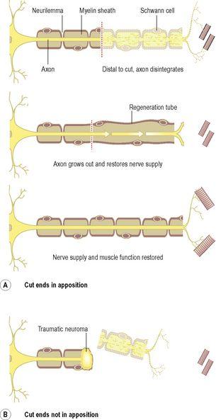
Figure 7.14
Regrowth of peripheral nerves following injury.
Neuroglial damage
Astrocytes
When severely damaged, astrocytes undergo necrosis and disintegrate. In less severe and chronic conditions there is proliferation of astrocyte processes and later cell atrophy (
gliosis
). This process occurs in many diseases and is analogous to fibrosis in other tissues.
Oligodendrocytes
These cells form and maintain myelin, having the same functions as Schwann cells in peripheral nerves. They increase in number around degenerating neurones and are destroyed in demyelinating diseases such as
multiple sclerosis
(
p. 178
).
Microglia
Microglia are thought to be derived from monocytes that migrate from the blood into the nervous system before birth, and are found mainly around blood vessels. Where there is inflammation and cell destruction the microglia increase in size and become phagocytic.
Central nervous system
The central nervous system consists of the brain and the spinal cord.
The meninges and cerebrospinal fluid (CSF)
Learning outcomes
After studying this section you should be able to:
describe the structure of the meninges
describe the flow of cerebrospinal fluid in the brain
list the functions of cerebrospinal fluid.
The meninges
The brain and spinal cord are completely surrounded by three layers of tissue, the
meninges
, lying between the skull and the brain, and between the vertebral foramina and the spinal cord. Named from outside inwards they are the:
•
dura mater
•
arachnoid mater
•
pia mater (
Fig. 7.15
).
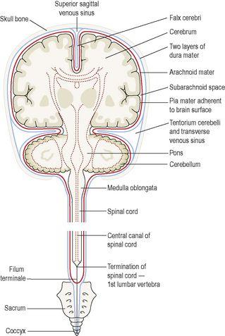
Figure 7.15
The meninges covering the brain and spinal cord.
The dura and arachnoid maters are separated by a potential space, the
subdural space
. The arachnoid and pia maters are separated by the
subarachnoid space
, containing
cerebrospinal fluid
.
Dura mater
The cerebral dura mater consists of two layers of dense fibrous tissue. The outer layer takes the place of the periosteum on the inner surface of the skull bones and the inner layer provides a protective covering for the brain. There is only a potential space between the two layers except where the inner layer sweeps inwards between the cerebral hemispheres to form the
falx cerebri
; between the cerebellar hemispheres to form the
falx cerebelli
; and between the cerebrum and cerebellum to form the
tentorium cerebelli
.
Venous blood from the brain drains into venous sinuses between the two layers of dura mater. The
superior sagittal sinus
is formed by the falx cerebri, and the tentorium cerebelli forms the
straight
and
transverse sinuses
(see
Figs 5.36
and
5.37
,
p. 97
).
Spinal dura mater forms a loose sheath round the spinal cord, extending from the foramen magnum to the 2nd sacral vertebra. Thereafter it encloses the
filum terminale
and fuses with the periosteum of the coccyx. It is an extension of the inner layer of cerebral dura mater and is separated from the periosteum of the vertebrae and ligaments within the neural canal by the
epidural space
(see
Fig. 7.27
), containing blood vessels and areolar connective tissue. It is attached to the foramen magnum and by strands of fibrous tissue to the posterior longitudinal ligament at intervals along its length. Nerves entering and leaving the spinal cord pass through the epidural space. These attachments stabilise the spinal cord in the neural canal. Dyes, used for diagnostic purposes, and local anaesthetics or analgesics to relieve pain, may be injected into the epidural space.
Arachnoid mater
This is a layer of fibrous tissue that lies between the dura and pia maters. It is separated from the dura mater by the
subdural space
, and from the pia mater by the
subarachnoid space
, containing
cerebrospinal fluid
. The arachnoid mater passes over the convolutions of the brain and accompanies the inner layer of dura mater in the formation of the falx cerebri, tentorium cerebelli and falx cerebelli. It continues downwards to envelop the spinal cord and ends by merging with the dura mater at the level of the 2nd sacral vertebra.
Pia mater
This is a delicate layer of connective tissue containing many minute blood vessels. It adheres to the brain, completely covering the convolutions and dipping into each fissure. It continues downwards surrounding the spinal cord. Beyond the end of the cord it continues as the
filum terminale
, pierces the arachnoid tube and goes on, with the dura mater, to fuse with the periosteum of the coccyx.
Ventricles of the brain and the cerebrospinal fluid
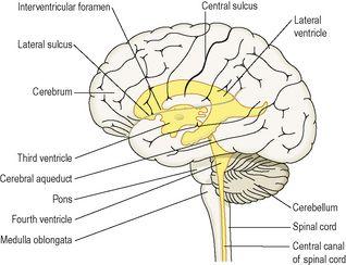
The brain contains four irregular-shaped cavities, or
ventricles
, containing
cerebrospinal fluid
(CSF) (
Fig. 7.16
). They are:
•
right and left lateral ventricles
•
third ventricle
•
fourth ventricle.


