Ross & Wilson Anatomy and Physiology in Health and Illness (202 page)
Read Ross & Wilson Anatomy and Physiology in Health and Illness Online
Authors: Anne Waugh,Allison Grant
Tags: #Medical, #Nursing, #General, #Anatomy

Gene mutation
Many diseases, such as cystic fibrosis (
p. 258
) and haemophilia (
p. 72
), are passed directly from parent to child via a faulty gene. Many of these genes have been located by mapping of the human genome, e.g. the gene for cystic fibrosis is carried on chromosome 7. Other diseases, e.g. asthma, some cancers and cardiovascular disease, have a genetic component (run in the family). In these cases, a single faulty gene has not been identified, and inheritance is not as predictable as when a single gene is responsible. The likelihood of an individual developing the disease depends not only on their genetic make-up, but also on the influence of other factors, such as lifestyle and environment.
Phenylketonuria
In this disorder, which is an example of an
inborn error of metabolism
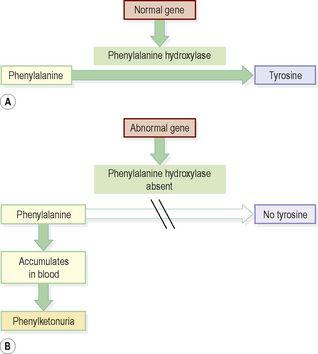
, the gene responsible for producing the enzyme phenylalanine hydroxylase is faulty, and the enzyme is absent. This enzyme normally converts phenylalanine to tyrosine in the liver, but in its absence phenylalanine accumulates in the liver and overflows into the blood (
Fig. 17.11
). In high quantities, phenylalanine is toxic to the central nervous system and, if untreated, results in brain damage and mental retardation within a few months. Because there are low levels of tyrosine, which is needed to make melanin, depigmentation occurs and children are fair skinned and blonde. The incidence of this disease is now low in developed countries because screening of newborn babies detects the condition and treatment is provided.
Figure 17.11
Phenylketonuria. A.
Gene function normal.
B.
Abnormal gene.
Chromosomal abnormalities
Sometimes during meiosis, a fault in the process produces a gamete carrying abnormal chromosomes – too many, too few, abnormally shaped, or with segments missing. Often, these aberrations are lethal and a pregnancy involving such a gamete miscarries in the early stages. Non-lethal conditions include Down syndrome and cri-du-chat syndrome.
Down syndrome
In this disorder, there are three copies of chromosome 21 (trisomy 21), meaning that an extra chromosome is present, caused by failure of chromosomes to separate normally during meiosis. People with Down syndrome are usually short of stature, with pronounced eyelid folds and flat, round faces. The tongue may be too large for the mouth and habitually protrudes. Learning disability is present, ranging from mild to severe. Life expectancy is shorter than normal, with a higher than average incidence of cardiovascular and respiratory disease. Down syndrome is associated with increasing maternal age, especially over 35 years.
Cri-du-chat syndrome
Cri-du-chat (cat’s cry) refers to the characteristic meowing cry of an affected child. This syndrome is caused when part of chromosome 5 is missing, and is associated with learning disabilities and anatomical abnormalities, including gastrointestinal and cardiovascular problems.
For a range of self-assessment exercises on the topics in this chapter, visit
www.rossandwilson.com
.
CHAPTER 18
The reproductive systems
Female reproductive system
438
External genitalia (vulva)
438
Internal genitalia
439
Vagina
439
Uterus
440
Uterine tubes
442
Ovaries
442
Puberty in the female
444
The reproductive cycle
444
Menopause
445
Breasts
446
Male reproductive system
447
Scrotum
447
Testes
448
Seminal vesicles
449
Ejaculatory ducts
449
Prostate gland
449
Urethra and penis
449
Ejaculation
450
Puberty in the male
450
Sexually transmitted infections
452
Diseases of the female reproductive system
453
Pelvic inflammatory disease (PID)
453
Vulvar dystrophies
453
Imperforate hymen
453
Disorders of the cervix
453
Disorders of the uterine body
453
Disorders of the uterine tubes and ovaries
454
Female infertility
454
Disorders of the breast
455
Diseases of the male reproductive system
455
Infections of the penis
455
Infections of the urethra
455
Epididymis and testes
456
Prostate gland
456
Breast
456
Male infertility
457
ANIMATIONS
18.1
Female external genitalia
438
18.2
Vestibular glands
439
18.3
Female reproductive ducts
439
18.4
Ovaries
442
18.5
Menstrual cycle
445
18.6
Ovarian cycle
445
18.7
Breasts
446
18.8
Testes
448
18.9
Spermatozoa
448
18.10
Male accessory sex glands
449
18.11
Male reproductive ducts
449
18.12
Male external genitalia
450
18.13
Pathway of sperm
450
The ability to reproduce is one of the properties distinguishing living from non-living matter. The more primitive the animal, the simpler the process of reproduction. In human beings the process is one of sexual reproduction, in which the male and female organs differ anatomically and physiologically, and the new individual develops from the fusion of two different sex cells (gametes).
Adult males and females produce specialised reproductive germ cells, called
gametes
. The male gametes are called
spermatozoa
and the female gametes are called
ova
. They contain the genetic material, or
genes
, on
chromosomes
, which pass inherited characteristics on to the next generation. Other body cells possess 46 chromosomes arranged in 23 pairs but the gametes contain only 23, one from each pair. Gametes are formed by
meiosis
(
p. 432
). At
fertilisation
, the fusion of an ovum and a spermatozoon, the resulting cell is called a
zygote
, and now possesses the full complement of 46 chromosomes.
The zygote embeds itself in the wall of the uterus where it grows and develops during the 40-week
gestation period
before birth.
The functions of the female reproductive system are:
•
formation of ova
•
reception of spermatozoa
•
provision of suitable environments for fertilisation and fetal development
•
parturition (childbirth)
•
lactation, the production of breast milk, which provides complete nourishment for the baby in its early life.
The functions of the male reproductive system are:
•
production of spermatozoa
•
transmission of spermatozoa to the female.
The later sections of this chapter describe disorders of the reproductive systems.
Female reproductive system


