Sexology of the Vaginal Orgasm (6 page)
Read Sexology of the Vaginal Orgasm Online
Authors: Karl F. Stifter

- A Solemn Oblation
- A Solemn Oblation
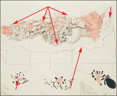
Vulva motives as votive offerings date back to ancient times. A 4,000-year-old bronze plate was found at an excavation site in ancient Babylon. Shaped like a pubic triangle it is dedicated to Ishtar, the goddess of life, and bears a moving inscription: “
When Sarrurn-ken was Lord of Assur, Adeturn, the wife of Belumnada, gave to Ishtar of Assur, her mistress, a votive offering. For the life of her husband, her own life and that of her child she brought a vulva to the temple.
” (Jakob-Rost, L.
& Freydank, H., 1981; p. 325-327)
Another patient donated to this goddess “a vulva made of lapis lazuli with a little star of gold” (Andrae, 1935; p. 36). What is striking when one reads this text that was written thousands of years ago is the distinctly positive image that was attributed to the vagina. For example, a lover enthuses about his girl: “
Her vagina, like her mouth, is sweet … a mouth of pleasure, a … mouth of honey.
” (Alster, 1985; 133: p.12-13)
At other places and at other times the symbolized female genitals were revered as a sign of immense power. The
Zuni
Indians in New Mexico, for example, carved it into the rock and worshipped it as the
Great Mother
. And in tantric Hin- duism the
Yoni Yantra
, the downward-pointing triangle, is the very archetype of femaleness.
5.
The
G Spot

- The Female Prostate
The female urethra, measuring 3 – 5 cm in length, forms an inseparable part of the front side of the vaginal wall facing the stomach. In evolutionary terms, it corresponds to the section of the male urethra which runs through the pros- tate. It thus does not come as a surprise that even the ureth- ra of the woman is surrounded by glands with outpouching ducts. These are referred to as paraurethral glands or also
Skene’s ducts
. This term was coined by the American gyne- cologist Skene who believed to have discovered these glands in 1880. He and his colleagues at the time obviously did not know that Herophile had already discovered this glandular formation as the “female prostate” in 300 B.C. All of the great doctors of the Middle Ages were also fami- liar with it. “I know nothing about the physiology of these glands...”, wrote Skene, “...what is their function is a question to be answered in the future” (Skene, 1880; p. 267). One hundred years had to pass before modern medicine also recognized that Herophile was right.In 1947 an imaginative researcher succeeded in drawing a vivid image of these glands for his contemporaries. The gynecologist J. W. Huffman pressed hot wax into the urethra of female corpses. He then presented 3-dimensio- nal wax models of the paraurethral glands at a meeting of American gynecologists.
Fig. 17 shows an enlargement of one of these exhibits. The
2-8 cm long urethra can be seen in red. On the right-hand side, one sees the outer opening of the urethra, and on the
prostative glands outer urethral opening
left-hand side the part located next to the blad- der.
Fig. 17
cross section
upper wall of vagina
Wax model of female prostate
The prostatic glands and ducts are depicted in white. On the base one sees the vaginal canal that runs paral- lel to the upper
vaginal wall. The small images below the model show cross- sections taken at three different locations. To enable better differentiation the richly furrowed urethra is shown in red and the surrounding ducts of the prostate are shown in black.
Fig. 18 shows a circular view of the corresponding passages leading into the urethra. Its outer end is visible at the bot- tom of the picture.
Fig. 18
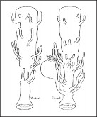
urethral ducts
The most surprising finding of Huffman’s study which was based on a total of eleven female corp- ses was certainly the size, the quantity and diversity of the paraurethral ducts. Thus these formations could not just be arranged all over the urethra but also primarily around the outer or inner urethral opening. Huffman
compares the female prostate with a tree. The urethra could be seen as a tree trunk and the outpouching ducts as race- mose or branch-like formations.
In his dissertation in 1985, Milan Zaviacˇicˇ who heads the Institute for Experimental Pathology in Bratislava proved that the female prostate is not merely an atrophied forma- tion but rather an organ with a highly complex secretion- related activity both inside and outside. In none of his stu- dies was he able to find an enzyme that was characteristic for the male prostate which he did not also find in the fe- male prostate tissue. (Zaviacˇicˇ , 1999)
The average weight of the female prostate in an adult woman is about 5 g and corresponds to a fifth to a fourth of the weight of an adult man’s prostate. It is about 3 cm long, 2 cm wide and 1 cm high. It corresponds to the male coun- terpart so much that it even shows the same illnesses which only appear much more rarely. This includes benign hyper- trophia and prostatitis and even the prostate carcinoma (Sesterhenn et al., 1998; Zaviacˇicˇ et al., 1993a).
The female prostate is thus not an atrophied organ but rath- er a functional urogenital one. Accordingly, it also plays an important sexological role, since prostatic tissue can be highly erogenous.
The sexual pleasure triggered by stimulation of the pros- tate is something many homosexual men take for granted. When they have this spot massaged by anal contact they often experience orgasms- sometimes even multiple orgasms.
It is thus obvious that climaxes can also be triggered by sui-
table vaginal stimulation of the female prostate and after adequate training.
- Gräfenberg and the Consequences
An article by Ernst Gräfenberg, dated 1950, gives us the first report about this highly sensitive spot on the front wall of the vagina.
The German gynecologist who was born on September 26, 1881 in Adelebsen near Göttingen, emigrated to America in 1940 where he died in 1957. He published a four-page article in
The International Journal of Sexology
on the role of the urethra in female orgasm. This contri- bution was to have unexpected consequences for modern sexology. Since this journal was published in Bombay of all places it was hardly accessible so that it fell into oblivion for thirty years. The original article has thus only been read by a few. This had lead to some fatal mis- understanding that I will attempt to do away with in the following.Gräfenberg wrote:
“An erotic zone always could be demon- strated on the anterior wall of the vagina along the course of the urethra ...Women tested this way always knew when the fin- ger slipped from the urethra by the impairment of their sexual stimulation. During orgasm this area is pressed downwards against the finger like a small cystocele protruding into the vaginal canal
” (Gräfenberg, 1950; p. 146)This passage clearly shows that Gräfenberg described the entire upper vaginal wall in which the 3 cm long urethra run, as a highly erogenous area. Then, however, a crucial
sentence follows: “…The most stimulating part is located at the posterior urethra, where it arises from the neck of the bladder.
” And it is precisely this part that John Perry and Beverly Whipple called, in his commemoration, the Gräfenberg spot.Accordingly, the abbreviated version of the title of their international bestseller was
The G spot
. This was formula- ted in a well-meaning but fatal way, since it reduced the entire vaginal area along the urethra to a point by referring to it as a “spot”. Subsequently, millions of couples tried to pinpoint the spot that was described as a “
pea-sized pleasure button
” and other similar expressions in pseudo-scientific publications and magazines.The most disappointing search failed for a different reason. The G spot authors claimed that the
G spot
was located 2 inches (5 centimeters) away from the vaginal opening (Ladas, Whipple, Perry, 1982). This, however, does not cor- respond with Gräfenberg’s information since the urethra generally only measures about 4 cm “
where it exits from the neck of the bladder
” to the opening. Thus, in anatomical terms, the crucial “
spot
” can also be located just as deeply in the vagina. This is clearly shown by fig. 19. If it were located any deeper then this would mean that the G spot would lie beyond the female prostatic glands.The misleading information (the spot located 5 cm deep) did not just lead to disappointment and doubts among cou- ples.
Another consequence has been that to this very day sexolo- gists are debating whether there is an autonomous pleasure center that exists independently of the female prostate.
G spot
uterus
bladder
Fig. 19
vaginal opening urethral opening
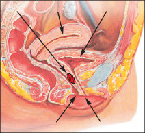
Location of G spot
Some even claim that the G spot is merely a myth (Hines, 2001). And in encyclopedias and official information pages in the Internet this “bean-shaped mass of nerve tissue” is simply imagined to be located some- where behind the pubic bone. In gui-
des to sex it is described as the “lower side of the clitoris”. (e.g., in: Bodansky Steve and Vera, 2002, p. 83)
This entire discussion is a silly quarrel over trifles, since the G spot is not an anatomical organ in the true sense of the word. It is the
imagined
area on the upper vaginal wall, behind which the prostatic glands are located along the urethra. When pressure is applied these glands swell and with greater familiarity and individual learning they are sexually highly excitable. In my view it is contrived but even so it can hardly be disputed in sexological terms.Accordingly, Gräfenberg also spoke of an erogenous “zone“ on the vaginal wall. By contrast, Beverly Whipple who coi- ned the term “G-spot” is not referring to a “zone” but uses this word synonymously with the female prostate. (Whip- ple 2005) A term designating a certain spot has thus come to describe an organ. The gland itself emerges from an area from which it can be stimulated vaginally. This ambiguity has given way to a number of misunderstandings. They have been additionally reinforced by misleading illustrations of
the G spot which is shown as being located in the same pla- ce in all women.
If Gräfenberg discovered that the most “stimulating part” of the erogenous, upper vaginal wall was located where the urethra “
exits the neck of the bladder
” then this shouldn’t be generalized and interpreted in a narrow-minded way for a number of reasons. First of all, one should bear in mind that his patients were in a prostrate position when he sti- mulated them with a finger. It can be assumed, he did this in a “sufficient” way from behind the pubic bone. Thus ero- ticising areas were automatically included that do not lie so deep inside the vagina. If for normal stimulation also the two finger tips of the middle and index finger are used, it is not possible from the outset to localize this area to the millimeter. The pressure surface on the upper vaginal wall is too large in comparison to the approximately 3 cm long path along the urethra to be able to say anything specific about differences in pleasure. Moreover, with increasing sti- mulation a swelling may produce in this area that reaches the size of a Euro coin.Huffman’s wax models have also shown that the female pros- tatic glands can differ greatly in position, shape and size around the urethra. For instance, Zaviacˇicˇ discovered that in 66% of the female autopsies the mentioned glands were mainly located around the outer opening of the urethra. (Zaviacˇicˇ & Ablin, 2002) It might be added that this moti- vated Edward Eichel (1977) to develop his ideas on the
Coi- tal Alignment Technique
(CAT) in which the front part of the female urethra is directly stimulated by pressure and counter pressure of the male and female genital regions. Recently, special ultrasonographic stuides have provided nteresting information on the location of the G spot.Fig. 20
Bladder Urethra Prostate Vagina

Ultrasound image of female prostate
These studies were filmed for the docu- mentary about the
PELflex
orgasm trai- ner in Vienna. (Stifter& Stackl,2002)
Fig. 20 depicts the prostate of a 45-year old woman. The gland structure runs like a blind tube below the
urethra, which has about 3/4 its length and practically shares the same opening. To date position and extension have remained largely unknown. Even Milan Zaviacˇicˇ who has seen more paraurethral glands than anyone else on earth, was greatly surprised by these shots. Detailed urological and gynecological examinations were able to ascertain that this picture did not depict a urethral protrusion (diverticle).
Bimholz (2001) has also used ultrasound technology to show that the inner legs of the clitoris run much deeper than was originally assumed and only end in the vicinity of the G spot. This could possibly indicate that the cavernous clitoral tissue is part of the structure of the G spot. The local swelling of this area, when stimulated the detumescence following orgasm, could thus not be explained in terms of the prostatic glands being filled with fluid. The findings by the Israeli sexologist Zvi Hoch (1980) also make it plausible that in some women the stimulation of the front vaginal wall could be even more exciting than a direct stimulation of the clitoris.
Other books
A Wild Goose Chase Christmas: Quilts of Love Series by Jennifer AlLee
The Enemy Inside by Steve Martini
The Winter Sea by Susanna Kearsley
Mr Hire's Engagement by Georges Simenon
Moon River by J. R. Rain
Come Home Bad Boy by Leah Holt
The Lion of Justice by Jean Plaidy
Rush by Nyrae Dawn
Unplanned: The Dramatic True Story of a Former Planned Parenthood Leader's Eye-Opening Journey Across the Life Line by Abby Johnson, Cindy Lambert