Pocket Medicine: The Massachusetts General Hospital Handbook of Internal Medicine (116 page)
Read Pocket Medicine: The Massachusetts General Hospital Handbook of Internal Medicine Online
Authors: Marc Sabatine
Tags: #Medical, #Internal Medicine

BOOK: Pocket Medicine: The Massachusetts General Hospital Handbook of Internal Medicine
7.9Mb size Format: txt, pdf, ePub
Initial treatment
• Resuscitation, control airway, monitor vital signs, fingerstick glucose, IV access
• Immobilization of C-spine if concern for cervical trauma
• Thiamine (100 mg IV)
prior to dextrose
to prevent exacerb. of Wernicke’s encephalopathy
• Dextrose (50 g IV push)
• Naloxone 0.01 mg/kg if opiates suspected; supportive care important in nearly all tox cases
• If concern for ↑ ICP ± herniation: ↑ head of bed; osmotherapy w/ mannitol or hypertonic saline; ↑ ventilation; dexamethasone for tumor edema; c/s neurosurgery (? decompress)
Diagnostic studies (
Continuum
2011;17:967)
• Labs: CBC, electrolytes, BUN/Cr, LFTs, NH
3
, tox screen, TSH, B
12
, ABG, U/A, ECG
• Imaging: head CT, consider MRI; radiographs to r/o C-spine fracture; CXR
• Lumbar puncture to r/o meningitis, SAH or noninfectious inflammation (eg, autoimmune)
• EEG to evaluate for nonconvulsive seizures, toxic/metabolic encephalopathy
Further treatment of delirium (
Annals
2011;154:746)
• Treat underlying acute illness, eliminate precipitating factors, provide supportive care
• Address sensory & cognitive impairments, increase familiarity
• Decrease/prevent infection/restraints if possible, remove lines/catheters if unnecessary
• Promote good sleep: reduce noise & night-time interventions; selective med if necessary
• Meds: consider antipsychotics, avoid benzos except for alcohol withdrawal or seizures
ANOXIC BRAIN INJURY
Prevalence (
NEJM
2012;367:1912)
• Pts with at least 5 min of cerebral hypoxia at risk • 1.5 million cardiac arrests per year in U.S.; for inPt arrest,
20% survival,
70% of Pts who survive will have a good long-term neurologic outcome
Initial evaluation (
Circulation
2010:S768)
• Neuro exam: arousal/verbal, eyes & other cranial nerves, motor response to pain • Imaging: usually not informative w/in first day after arrest, but should be done prior to initiating hypothermia if patient found down or has had head trauma
Induced hypothermia (
Circulation
2008;118:2452 & 2013;127:244)
• Indications: comatose (eg, no meaningful response to verbal stimuli) <6 h following cardiac arrest (not isolated resp. arrest). Fully studied only in VT/VF, but consider after asystole or PEA arrest or 6–12 h after cardiac arrest.
• Exclusion: pregnancy, CV instability despite pressors/assist devices, other cause of coma, persistent ↓ O
2
• Relative contraindications: major head trauma, coagulopathy/bleeding, major surgery <14 d, systemic infection/sepsis • Method: target temp 32–34°C × 24 h (from time of initiation of cooling). Can use cold saline infusions; ice packs to the head, neck and torso; cooling blankets; cooling vest or endovascular catheter if available. Goal to achieve target temp <6 h. Start rewarming 24 h after cooling is initiated (rewarm no faster than 0.5°C per h).
• Complications
cardiac dysrhythmias (bradycardia most common): if signif dysrhythmia or hemodynamic instability, d/c cooling and rewarm patient
coagulopathy: Pts can receive fibrinolytics, GP IIb/IIIa inhibitors, etc., and still undergo cooling. ✓ PT and PTT.
infection: ✓ surveillance blood cultures during cooling
hyperglycemia during cooling, hypoglycemia w/ rewarming; stop insulin if glc <200 mg/dL
hypokalemia during cooling, hyperkalemia w/ rewarming; keep K 4–5 mEq/L
Ongoing evaluation
• Neuro exam: daily focus on coma exam. No exam finding is reliable <24 h or on sedation. Pt needs to be off sedation for an adequate time to evaluate (depends on doses used, duration of Rx, metabolic processes in the individual Pt).
• Labs: daily CBC, PT/PTT, electrolytes. Serum neuron-specific enolase (NSE) on days 1–3
• Imaging: noncontrast CT 24 h after arrest; if unrevealing, consider MRI around days 3–5
• EEG: consider in all to exclude seizures or myoclonus; greatest risk during rewarming • Somatosensory evoked potentials (SSEP): helpful for prediction of poor outcome if cortical responses are absent bilaterally; perform 48 h after arrest (72 h if cooled)
Prognosis (
Neuro
2006;67:203;
NEJM
2009;361:605)
• Prior to cooling era, uniformly poor prognosis could be predicted at 72 h only in Pts who have absent pupillary and corneal reflexes, and no motor response to pain; or with absent SSEPs at 48 h. With cooling, it is less clear if the prior measures are as reliable.
• Otherwise, prognosis requires multifactorial approach considering exam, age, comorbid diseases, ancillary data (NSE, EEG, SSEP; imaging is less reliable for poor outcome) • When in doubt, err on the side of giving more time (esp. in younger Pts and induced hypothermia Pts)
SEIZURES
Definitions (
NEJM
2003;349:1257;
Epilepsia
2010;51:676)
•
Seizure
= abnormal, paroxysmal, excessive discharge of CNS neurons; occurs in 5–10% of the population; can range clinically from dramatic to subtle •
Epilepsy
= recurrent unprovoked seizures; 0.5–1.0% of population •
Generalized seizures
(involves brain diffusely)
Tonic-clonic
(grand mal): tonic phase (10–20 sec) with contraction of muscles (causing expiratory moan, cyanosis, pooling of secretions, tongue biting) → clonic phase (~30 sec) with intermittent relaxing and tensing of muscles
Absence
(petit mal): transient lapse of consciousness w/o loss of postural tone, usu pedi
Myoclonic
(infantile spasms & juvenile myoclonic epilepsy): sudden, brief contraction
•
Focal (partial) seizures
(involves discrete brain area, implies a structural lesion)
Simple
(w/o Δ MS) vs.
complex
(w/ Δ MS): motor, sensory and/or autonomic
Focal with secondary generalization:
starts focal, becomes generalized
Differential diagnosis
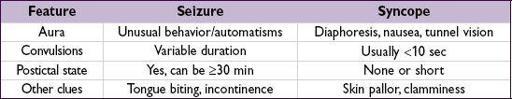
•
Syncope
(
Lancet Neurol
2006;5:171)
•
Nonepileptic seizure
(NES, aka “psychogenic”): may see side-to-side head turning, asymmetric large-amplitude limb movements, diffuse shaking w/o LOC, and crying or talking during event • Other: metabolic disorders (eg, alcoholic blackouts, hypoglycemia), migraine, TIA, transient global amnesia, narcolepsy (cataplexy), nonepileptic myoclonus, tics, asterixis
Etiologies (varies strongly by age)
•
A
lcohol withdrawal, illicit drugs, meds (eg, β-lactams, bupropion, tramadol, metronidazole, meperidine, CsA, antidep., clozapine can lower seizure threshold) •
B
rain tumor or penetrating trauma •
C
erebrovascular disease, including subdural hematomas, hypertensive encephalopathy •
D
egenerative disorders of the CNS (eg, Alzheimer’s) •
E
lectrolyte (hyponatremia) & other metabolic (eg, uremia, liver failure, hypoglycemia) • Idiopathic (in ~60%)
Clinical manifestations
•
Aura
(sec to mins): premonition with paresthesias, focal motor contractions, abnormal smells/tastes, fear, depersonalization, déjà vu, autonomic changes, automatisms •
Ictal period
(sec to mins): tonic and/or clonic movements of head, eyes, trunk or extrem.
•
Postictal period
(mins to h): slowly resolving period of confusion, disorientation, and lethargy. May be accompanied by focal neurologic deficits (Todd’s paralysis).
•
Status epilepticus
: continuous tonic-clonic seizure ≥30 min or repeated seizures w/o resolution of postictal encephalopathy. Complications include neuronal death, rhabdomyolysis and lactic acidosis.
•
Nonconvulsive status epilepticus
: alteration of awareness (ranging from confusion to coma) w/o motor manifestations of seizure. Dx with EEG.
Clinical evaluation
• Seizure: patient usually w/o recollection, must talk to witnesses
unusual behavior before seizure (ie, an aura)
type & pattern of abnl movements, incl. head turning & eye deviation (gaze preference usually
away
from seizure focus)
loss of responsiveness
• HPI: recent illnesses/fevers, head trauma, sleep deprivation, medication compliance • PMH: prior seizures orFHx, prior meningitis/encephalitis, prior stroke or head trauma • Medications, alcohol and illicit drug use • General physical exam should include the skin, looking for neuroectodermal disorders (eg, neurofibromatosis, tuberous sclerosis) that are a/w seizures • Neurologic exam should look for focal abnormalities → underlying structural abnormality
Diagnostic studies (
Neurology
2007;69:1996)
• Laboratory: full electrolytes, BUN, Cr, glc, LFTs, tox screen, medication levels • EEG: during seizure can capture repetitive rhythmic activity (generalized seizures will typically have abnl EEG; partial may not); interictal EEG normal in 50% of Pts w/ epilepsy, and interictal epileptiform activity (spikes or sharp waves) seen in only 25% of Pts w/ epilepsy but up to 2% of normal population; sleep deprivation and repeated studies ↑ dx yield of EEG; video monitoring may help w/ nonepileptic seizures • MRI to r/o structural abnormalities; ↑ Se w/ fine coronal imaging of frontal & temporal lobes • LP (if no space-occupying lesion on imaging): if suspect meningitis (eg, fever, ↑ WBC, nuchal rigidity) or encephalitis and in
all
HIV
Treatment (
Lancet
2006;367:1087 & 2007;369:1000, 1016;
NEJM
2008;359:166)
Other books
The Wayward One (The De Montforte Brothers Book 5) by Danelle Harmon
The List (Zombie Ocean Book 5) by Michael John Grist
Life For a Life by T F Muir
Untimely You by K Webster
Finding Midnight by T. Lynne Tolles
Ask No Questions by Elyot, Justine
(1964) The Man by Irving Wallace
The Last of the Gullivers by Carter Crocker