i bc27f85be50b71b1 (4 page)
Read i bc27f85be50b71b1 Online
Authors: Unknown

1
Cardiac System
Sean M. Collins
Introduction
Physical therapists in acute care faci lities commonly encounter patients
with cardiac system dysfunction as either a primary morbidity or
comorbidity. Based on current estimates, 59,700,000 Americans have
one or more types of cardiovascular disease (CVD), making the prevalence rate for men and women 20% ' In 1 997, CVD ranked first among all disease categories and accounted for 6, 1 45,000 inpatient
admissions.' In the acute care setting, the role of the physical therapist
with this diverse group of patients remains founded in examination,
evaluation, intervention, and discharge planning, for the purpose of
improving functional capacity and minimizing disabiliry. The physical
therapist must be prepared to safely accommodate for the effects of
dynamic (pathologic, physiologic, medical, and surgical intervention)
changes into his or her evaluation and intervention.
The normal cardiovascular system provides the necessary pumping
force to circulare blood through the coronary, pulmonary, cerebral,
and systemic circulation. To perform work, such as during functional
rasks, energy demands of the body increase, therefore increasing the
oxygen demands of the heart. A variery of pathologic states can create

2
Acme CARE HANDBOOK FOR PHYSICAL THERAPISTS
impairments in the cardiac system's ability to successfully meet these
demands, ultimately leading to functional limitations. To fully
address these functional limitations, the physical therapist must
understand normal and abnormal cardiac function, clinical tests, and
medical and surgical management of the cardiovascular system.
The objectives of this chapter are to provide the following:
1 . A brief overview of the Structure and function of the cardio-
vascular system
2.
An overview of cardiac evaluation, including physical exami-
nation and diagnostic testing
3.
A description of cardiac diseases and disorders, including
clinical findings and medical and surgical management
4.
A framework on which to base physical therapy evaluation
and intervention in patienrs with CVD
Structure
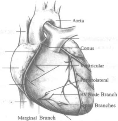
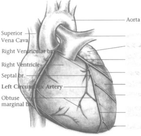
The heart and the roots of the great vessels ( Figme 1 - 1 ) occupy the pericardium, which is located in the mediastinum. The sternum, the costal cartilages, and the medial ends of the third to fifth ribs on the left side of
the thorax create the anterior border of the mediastinum. It is bordered
inferiorly by the diaphragm, posteriorly by the vertebral column and ribs,
and laterally by the pleural cavity (which contains the lungs).' Specific
cardiac structures and vessels and their respective functions are outlined
in Tables 1 - 1 and 1-2.
Note: The mediastinum and the heart can be displaced from their
normal positions with changes in the lungs secondary to various disorders. For example, a tension pneumothorax will shift the mediastinum away from the side of dysfunction (see Chapter 2 for further description of pneumothorax).
Function
The cardiovascular system must adjust the amount of nutrient and oxygen rich blood pumped out of the heart (cardiac output [CO]) to meet the wide spectrum of daily energy (metabolic) demands of the body.


CARDIAC SYSTEM
3
Superior Vena ClIVI
Pulm��", Art,,,,
Branch
Sinus Node Branch
rush'
Branches
"n'''''
Branch
Right Coronary
Art,,>,
Inferior Vena CO'"
PostcnOf Dcscmdlng
Art,,,,
Pulmonary Artery
Left Atrium
Ldt Main Coronary
Artery
lsI Diagonal Branch
Left Venuicle
2nd Diagonal Branch
Left Anterior
Descending Artery
Apical Branches
Figure 1-1. Anatomy of the right coronary artery and Jeft coro,wry artery,
including left maiu, left auterior descending, and left circumflex coronary
arteries. (Reprinted with permissioll (rom R C Becker. Chest Pain: The Most
Common Complaints Series. Boston: Butterworth-Heinemann, 2000;26-28.)
The heart's ability to pump blood depends on the following
characteristics3:
• Automaticity-the ability to initiate its own electrical
impulse
•
Excitability-the ability to respond to electrical stimulus

4 AClJTE CARE HANDBOOK FOR PHYSICAL THERAPISTS
Table 1-1. Primary Structures of the Hearr
Structure
Description
Function
Pericardium
Double-walled sac of elas
Prorects against infection
tic connective tissue, a
and trauma
fibrous outer layer and
serous inner layer
Epicardium
Outermost layer of car
Protects against infection and