In Search of Memory: The Emergence of a New Science of Mind (16 page)
Read In Search of Memory: The Emergence of a New Science of Mind Online
Authors: Eric R. Kandel
Tags: #Psychology, #Cognitive Psychology & Cognition, #Cognitive Psychology

IN THE LATE 1930S WADE MARSHALL WAS PROBABLY THE MOST
promising and accomplished young scientist working on the brain in the United States (figure 7–2). In a now classic series of studies he asked: How are the touch receptors of the body surface—the hands, face, chest, back—represented in the brain of cats and monkeys? Marshall and his colleagues discovered that the internal representation of touch is organized spatially: neighboring areas on the body surface are preserved in the brain.
By the time Marshall began his research, a great deal was already known about the anatomy of the cerebral cortex. The cortex is a convoluted structure that covers the two symmetrical hemispheres of the forebrain and is divided into four parts, or lobes (frontal, parietal, temporal, and occipital) (figure 7–3). Unfolded, the human cerebral cortex is about the size of a large cloth dinner napkin, only somewhat thicker. It contains about 100 billion neurons, each with about a thousand synapses, making a total of about 1 quadrillion synaptic connections.
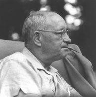
7–2
Wade Marshall (1907–1972) was the first scientist to map the detailed sensory representation of touch and vision in the cerebral cortex. He moved to the NIH in 1947 and became head of the Laboratory of Neurophysiology at NIMH in 1950, where I worked for him from 1957 to 1960. (Courtesy of Louise Marshall.)
Marshall began his studies of touch sensation as a graduate student at the University of Chicago in 1936. He discovered that moving the hairs on a cat’s leg or touching its skin produces an electrical response in specific groups of neurons in the somatosensory cortex, a region of the parietal lobe that governs the sense of touch. These studies showed only that the sense of touch is represented in the brain, but Marshall immediately realized that he could advance his analysis much further. He wanted to know whether neighboring areas of the skin are represented in neighboring areas of the somatosensory cortex or scattered at random across it.
For guidance in answering that question, Marshall sought postdoctoral training under Philip Bard, chairman of the department of physiology at the Johns Hopkins Medical School and a major figure in American biology. Marshall joined Bard in carrying out studies on monkeys in which they discovered that the entire body surface is represented in the somatosensory cortex in the form of a point-for-point neural map. Parts of the body surface that are adjacent to one another, such as the fingers, are represented near each other in the somatosensory cortex. A few years later a remarkably gifted Canadian neurosurgeon, Wilder Penfield, extended the study from monkeys to people, revealing that the parts of the body surface most sensitive to touch are represented by the largest areas of the somatosensory cortex (figure 7–4).
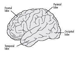
7–3 The four lobes of the cerebral cortex.
The frontal lobe is part of the neural circuit governing social judgments, planning and organization of activities, aspects of language, control of movement, and a form of short-term memory called working memory. The parietal lobe receives sensory information about touch, pressure, and space around the body and helps integrate that information into coherent perceptions. The occipital lobe is involved with vision. The temporal lobe is involved with auditory processing and aspects of language and memory.
Next, Marshall found that the light receptors in the retina of the eye are also represented in an orderly way in the primary visual cortex, a region of the occipital lobe. Finally, Marshall showed that the temporal lobe has a sensory map for sound frequencies, with different pitches represented systematically in the brain.
These studies revolutionized our understanding of how sensory information is organized and represented in the brain. Marshall showed that, even though the different sensory systems carry different types of information and end up in different regions of the cerebral cortex, they share a common logic in their organization: all sensory information is organized topographically in the brain in the form of precise maps of the body’s sensory receptors, such as, the retina of the eye, the basilar membrane in the ear, or the skin on the body surface.
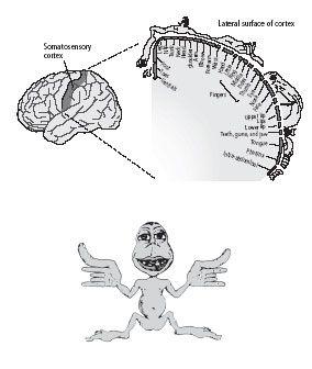
7–4 A sensory map of the body, as represented in the brain.
The somatosensory cortex—a strip in the parietal lobe of the cerebral cortex—receives sensations of touch. Each part of the body is represented separately. Fingers, mouth, and other particularly sensitive areas take up most space. Wilder Penfield called this cross-sectional map a sensory homunculus. A composite representation of the sensory homunculus as a figurine drawing (below) is based on the cross-sectional map. It shows a human with large hands, fingers, and mouth. (From
Mechanics of the Mind
, Colin Blakemore. © Cambridge University Press, 1977.)
These sensory maps are most easily understood by the representation of touch in the somatosensory cortex. Touch begins with receptors in the skin that translate the energy of a stimulus—for example, the energy transmitted by a pinch—into electrical signals in sensory neurons. The signals then travel along precise pathways to the brain, passing through several processing, or relay, stages in the brain stem and thalamus before terminating in the somatosensory cortex. At each stage the signals traveling from adjacent points on the skin are carried by nerve fibers that run alongside each other. In this way stimulation of two adjacent fingers, for instance, activates adjacent populations of nerve cells in the brain.
Knowledge of the brain’s sensory maps and an understanding of how they are organized topographically is exceedingly helpful in treating patients. Because these maps are incredibly precise, clinical neurology has long been an accurate diagnostic discipline, even though it has relied, until the recent development of brain imaging, only on the simplest, most primitive tools—a wad of cotton to test for touch, a safety pin to test for pain, a tuning fork to test for vibration, and a hammer to test reflex actions. Disturbances in the sensory and motor systems can be located with remarkable accuracy because of the one-to-one relationship between sites on the body and areas of the brain.
A dramatic example of this relationship is the Jacksonian sensory march, which characterizes a certain type of epileptic seizure first described by British neurologist John Hughlings Jackson in 1878. In such a seizure, numbness and sensations such as burning or prickling begin in one place and spread throughout the body. For example, numbness might begin at the fingertips and spread over the next minute to the hand, up the arm, across the shoulder, into the back, and down the leg on the same side. This sequence of sensations is explained by the arrangement of the sensory map of the body: the seizure, which is a wave of abnormal electrical activity in the brain, starts in the lateral area of the somatosensory cortex, where the hand is represented, and propagates across the cortex toward the midline, where the leg is represented.
Marshall’s marvelous scientific achievements came at a price, however. The experiments were physically demanding, often lasting more than twenty-four hours at a time. Frequently deprived of sleep, he became exhausted. In addition, there was tension with Bard. In 1942 Marshall collapsed with an acute psychotic paranoid episode after having actually threatened Bard physically. This episode of mental illness required that Marshall be hospitalized for eighteen months.
When Marshall returned to neuroscience in the late 1940s, he moved to a completely new set of problems: spreading cortical depression, an experimentally induced, reversible silencing of electrical activity in the cerebral cortex. By the time I arrived at NIH, he had passed the peak of his brilliant career. He still enjoyed doing occasional experiments, but he had lost his scientific drive and the clarity of his vision, and he focused much of his energy and interest on administrative matters, which he did well.
Although eccentric, moody, and somewhat suspicious in unpredictable ways, Marshall was a generous lab chief who was extremely supportive of the young people for whom he was responsible. I learned a great deal from him about the modesty and rigor that belong in a scientific laboratory. He had high standards of scientific conduct and a fine sense of humor about himself, which was reflected in the wonderful aphorisms he would call up at appropriate moments. One of his favorites, used whenever one of his findings was challenged, was, “We were confused and they were confused, but we were more accustomed to being confused.” On other occasions he would mutter, “Things will go along like this for a while and then they’ll get worse!”
In addition to humility, I learned from Marshall that with great personal strength and the passage of time, one might substantially recover from a severe mental illness (therapeutic drugs were not yet available). I also learned how much a person who had recovered from such a devastating disorder could accomplish. Many young people who went on to have superb careers in science and I myself owe our start and much of our later success to the personal and professional example of Wade Marshall. Despite my obvious inexperience, he did not insist that I work only on problems that interested him. Rather, he allowed me to think about what I wanted to do—which was to study how learning and memory are achieved in the cells of the brain. Science gives one a structured opportunity to try out ideas—and, if one is not afraid of falling on one’s face, to try out ideas that are raw, important, and bold. Marshall gave me the freedom to try to think creatively.
Grundfest, Purpura, Crain, Marshall, and later Steve Kuffler influenced me greatly. They transformed my life. They and Mr. Campagna, who paved the way for me to go to Harvard, illustrate the importance of student–teacher relationships in one’s intellectual development. Equally, they underscore the role of chance influences and of the generosity of spirit that encourages young people to thrive. Young people, for their part, must strive to have an open mind and seek out places where they will be surrounded by first-rate intellects.
B
y the time I arrived at Wade Marshall’s laboratory, I had progressed from the naïve notion of trying to find the ego, id, and superego in the brain to the slightly less vague idea that finding the biological basis of memory might be an effective approach to understanding higher mental processes. It was clear to me that learning and memory are central to psychoanalysis and psychotherapy. After all, aspects of many psychological problems are learned, and psychoanalysis is based on the principle that what is learned can be unlearned. In a large sense, learning and memory are central to our very identity. They make us who we are.
At that time, however, the biology of learning and memory was in a muddle. It was dominated by the thinking of Karl Lashley, a professor of psychology at Harvard who had convinced many scientists that there were no specific areas within the cerebral cortex that were specialized for memory.
Shortly after I arrived at NIMH, two research scientists changed all that. In a newly published research paper, Brenda Milner, a psychologist at the Montreal Neurological Institute of McGill University, and William Scoville, a neurosurgeon in Hartford, Connecticut, reported that they had tracked memory to specific regions in the brain. This news had a powerful impact on me and on many others, for it meant that perhaps there was at last a decisive end to a very old controversy about the human mind.
UNTIL THE MIDDLE OF THE TWENTIETH CENTURY, THE SEARCH
for where memory resides in the brain was shaped by two competing views of how the brain—and especially the cerebral cortex—works. One held that the cerebral cortex is composed of discrete regions with specific functions—one representing language, another vision, and so on. The other view was that mental capacities are products of the combined activity of the entire cerebral cortex.
The first person to champion the idea that particular mental abilities are located in specific regions of the cortex was Franz Joseph Gall, a German physician and neuroanatomist who taught at the University of Vienna from 1781 to 1802. Gall made two enduring conceptual contributions to the science of mind. First, he argued that
all
mental processes are biological, and so arise from the brain. Second, he proposed that the cerebral cortex has many distinct regions that govern specific mental functions.
Gall’s theory that all mental processes are biological put him at odds with dualism, the dominant theory of his time. Promulgated in 1632 by René Descartes, a mathematician and the father of modern philosophy, this theory holds that human beings have a dual nature: the body, which is material, and the soul, which resides outside the body and is immaterial and indestructible. This dual nature reflects two types of substances. The
res externa
—the physical substance that fills the body, including the brain—runs through the nerves and imbues the muscles with animal spirits. The
res cogitans
—the nonphysical stuff of thinking—is uniquely human. It gives rise to rational thought and consciousness, and it reflects in its nonphysical character the spiritual nature of the soul. Reflex actions and many other physical behaviors are carried out by the brain, while mental processes are carried out by the soul. Descartes believed these two agents interacted through the pineal gland, a small structure located deep in the middle of the brain.
The Roman Catholic Church, feeling its authority threatened by new discoveries in anatomy, embraced dualism because it separated the realms of science and religion. Gall’s radical argument for a materialist view of mind appealed to the scientific community because it ended the concept of the nonbiological soul, but it threatened the powerful conservative elements of society. Indeed, Emperor Francis I forbade Gall from lecturing in public and expelled him from Austria.
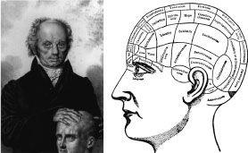
8–1 Phrenology
. Franz Joseph Gall (1758–1828) assigned different mental functions to specific regions of the brain, based on his observations. Gall later developed phrenology, a system that related personality to bumps on the skull. (Gall image courtesy of Anthony A. Walsh.)
Gall also speculated on which areas of the cortex do different things. Academic psychology at the time had settled on twenty-seven mental faculties. Gall assigned these faculties to twenty-seven different regions of the cortex, each of which he called a “mental organ.” (Additional regions were added later by Gall and others.) These mental faculties—such as factual memory, cautiousness, secretiveness, hope, belief in God, sublimity, parental love, and romantic love—were both abstract and complex, yet Gall insisted that each was controlled by a single, distinct region of the brain. This theory of localized function opened a debate that persisted through the next century.
Although correct in principle, Gall’s theory was flawed in its details. First, most of the “faculties” considered discrete mental functions in Gall’s time are far too complex to arise from single regions of the cerebral cortex. Second, Gall’s method of assigning functions to specific areas of the brain was misguided.
Gall distrusted studies of the behavior of people who had lost parts of their brain, so he ignored clinical findings. Instead, he developed an approach based on studies of the skull. He believed that each area of the cerebral cortex grew with usage and that this growth caused the overlying skull to protrude (figure 8–1).
Gall developed his idea in stages, beginning when he was young. In school, he had formed the impression that his most intelligent classmates had prominent foreheads and eyes. In contrast, a very romantic and enchanting widow he encountered had a prominent back of the head. Thus, Gall came to believe that great intelligence creates greater mass in the front of the brain, whereas great romantic passion produces greater mass in the back. In each case the overlying skull was enlarged by growth of the brain. Gall believed that by examining the bumps and ridges on the skulls of people well endowed with specific faculties, he could identify the centers of those faculties.
He systematized his thinking further when, as a young physician, he was put in charge of an insane asylum in Vienna. There he examined the skulls of criminals and found a bump above the ear that was remarkably similar to one found in carnivorous animals; Gall associated the bump with a part of the brain he believed was responsible for sadistic and destructive behavior. This approach to identifying the sites of mental faculties led to phrenology, a discipline that correlated personality and character with the shape of the skull.
By the late 1820s Gall’s ideas and the discipline of phrenology had become extremely popular, even among the public. Pierre Flourens, a French experimental neurologist, decided to put them to the test. Using various animals for his experiments, Flourens removed, one by one, the areas of the cerebral cortex that Gall had associated with specific mental functions, but he failed to find any of the behavioral deficits Gall had predicted. In fact, Flourens was unable to associate any deficits in behavior with specific regions of the cortex. It was only the size of the area removed, not its location or the complexity of behavior involved, that mattered.
Flourens therefore concluded that all regions of the cerebral hemispheres were equally important. The cortex is equipotential, he argued, meaning that every region is able to perform any of the brain’s functions. Thus, an injury to a particular region of the cerebral cortex would not affect one capacity more than another. “All perceptions, all volitions occupy the same seat in these [cerebral] organs; the faculty of perceiving, of conceiving, of willing merely constitutes thereby a faculty which is essentially one,” Flourens wrote.
Flourens’s views spread rapidly. Their swift acceptance was certainly due in part to the credibility of his experimental work, but it also represented a religious and political reaction against Gall’s materialistic view of the brain. If the materialistic view was correct, there was no need to postulate a soul as a necessary mediator of human cognitive functions.
THE DEBATE BETWEEN THE FOLLOWERS OF GALL AND THE
followers of Flourens colored thinking about the brain over the next several decades. It was not resolved until the second half of the nineteenth century, when the question attracted the attention of two neurologists, Pierre-Paul Broca in Paris and Carl Wernicke in Breslau, Germany. In the course of their studies of patients with specific language deficits, or aphasias, Broca and Wernicke made several important discoveries. Taken together, these discoveries form one of the most exciting chapters in the study of human behavior—namely, the first insight into the biological basis of a complex cognitive ability, language.
Rather than exploring the normal brain to test Gall’s ideas, as Flourens had done, Broca and Wernicke studied disease states—what physicians at that time referred to as nature’s experiments. They succeeded in associating specific disorders of language with damage to particular areas of the cerebral cortex, thus providing convincing evidence that at least some higher mental functions arise there.
The cerebral cortex has two important characteristics. First, although its two hemispheres appear to be mirror images of each other, they differ in both structure and function. Second, each hemisphere is concerned primarily with sensing and moving the opposite side of the body. Thus, sensory information that arrives at the spinal cord from the left side of the body—from the left hand, say—crosses over to the right side of the nervous system on its way to the cerebral cortex. Similarly, motor areas in the right hemisphere control movements of the left half of the body.
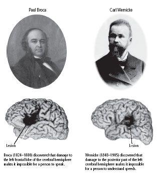
8–2 Two pioneers in the study of brain function in language.
(Portraits reprinted from
Essentials of Neural Science and Behavior
, Kandel, Schwartz, and Jessell, McGraw-Hill, 1995. Brain images courtesy of Hanna Damasio.)
Broca (figure 8–2), who was also a surgeon and anthropologist, founded what we now call neuropsychology, a science that examines alterations in mental processes produced by brain damage. In 1861 he described a fifty-one-year-old Parisian shoemaker named Leborgne who had suffered a stroke twenty-one years earlier. As a result of his stroke, Leborgne had lost the ability to speak fluently, although he indicated with facial expressions and actions that he understood the spoken language quite well. Leborgne had none of the conventional motor deficits that would affect speech. He had no difficulty moving his tongue, mouth, or vocal cords. In fact, he could utter isolated words, whistle, and sing a melody without difficulty, but he could not speak grammatically or create complete sentences. Moreover, his difficulty was not limited to the spoken language; Leborgne could not express his ideas in writing either.
Leborgne died one week after Broca first examined him. At the postmortem examination Broca discovered a damaged area, or lesion, in a region of the frontal lobe now called Broca’s area (figure 8–2). He went on to study, after their deaths, the brains of eight other patients who had been unable to speak. Each had a similar lesion in the frontal lobe of the left cerebral hemisphere. Broca’s findings provided the first empirical evidence that a well-defined mental capacity could be assigned to a specific region of the cortex. Since all the patients’ lesions were in the left hemisphere, Broca established that the two hemispheres, though apparently symmetrical, have different roles. This discovery led him to announce, in 1864, one of the most famous principles of brain function:
“Nous parlons avec l’hémisphère gauche!”
(We speak with the left hemisphere!)
Broca’s discovery stimulated a search for the location of other behavioral functions in the cortex. Nine years later two German physiologists, Gustav Theodor Fritsch and Eduard Hitzig, galvanized the scientific community when they showed that dogs would move their limbs in predictable ways when a specific region of the cerebral cortex was stimulated electrically. Moreover, Fritsch and Hitzig identified the small areas of the cortex that controlled the individual muscle groups responsible for the movements.